Prieto Sandra P, Powless Amy J, Boice Jackson W, Sharma Shree G, Muldoon Timothy J
Biomedical Engineering Department, University of Arkansas, Fayetteville, Arkansas 72701, United States of America.
10810 Executive Center Dr., Nephropath Ste. 100, Little Rock, Arkansas 72211, United States of America.
PLoS One. 2015 May 11;10(5):e0125598. doi: 10.1371/journal.pone.0125598. eCollection 2015.
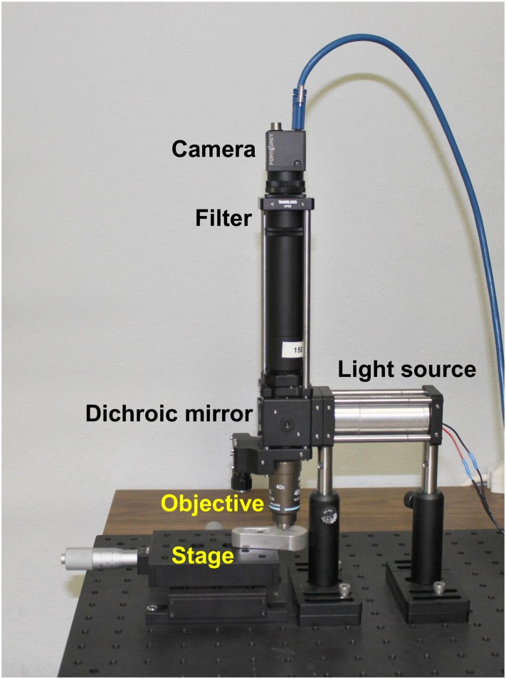
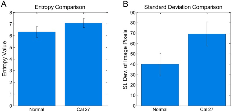
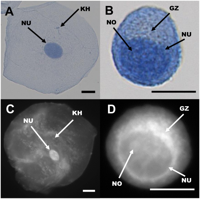
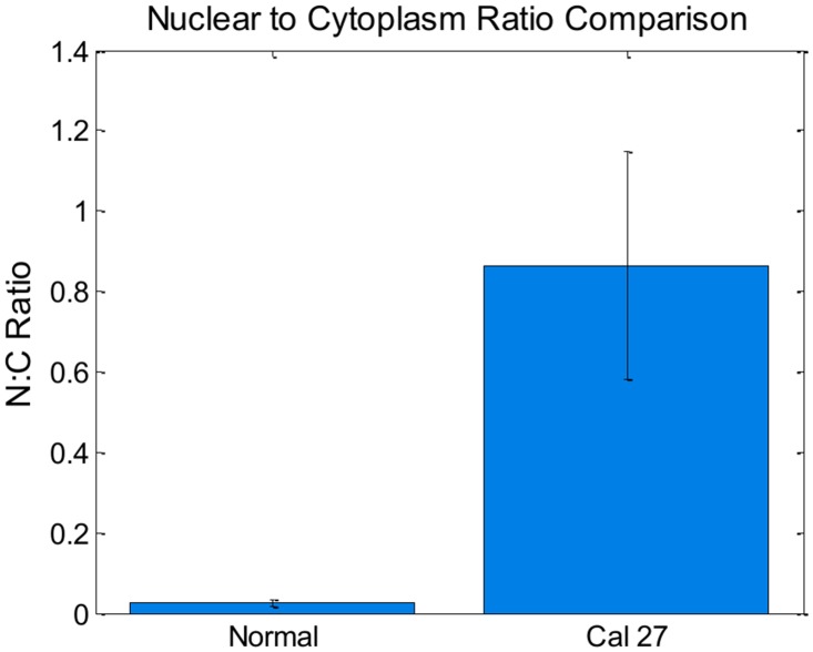
Proflavine hemisulfate, an acridine-derived fluorescent dye, can be used as a rapid stain for cytologic examination of biological specimens. Proflavine fluorescently stains cell nuclei and cytoplasmic structures, owing to its small amphipathic structure and ability to intercalate DNA. In this manuscript, we demonstrated the use of proflavine as a rapid cytologic dye on a number of specimens, including normal exfoliated oral squamous cells, cultured human oral squamous carcinoma cells, and leukocytes derived from whole blood specimens using a custom-built, portable, LED-illuminated fluorescence microscope. No incubation time was needed after suspending cells in 0.01% (w/v) proflavine diluted in saline. Images of proflavine stained oral cells had clearly visible nuclei as well as granular cytoplasm, while stained leukocytes exhibited bright nuclei, and highlighted the multilobar nature of nuclei in neutrophils. We also demonstrated the utility of quantitative analysis of digital images of proflavine stained cells, which can be used to detect significant morphological differences between different cell types. Proflavine stained oral cells have well-defined nuclei and cell membranes which allowed for quantitative analysis of nuclear to cytoplasmic ratios, as well as image texture analysis to extract quantitative image features.
硫酸普罗黄素是一种吖啶衍生的荧光染料,可作为生物标本细胞学检查的快速染色剂。由于其两亲性结构小且具有嵌入DNA的能力,普罗黄素能对细胞核和细胞质结构进行荧光染色。在本论文中,我们使用定制的便携式LED照明荧光显微镜,展示了普罗黄素作为快速细胞学染料在多种标本上的应用,这些标本包括正常脱落的口腔鳞状细胞、培养的人口腔鳞状癌细胞以及来自全血标本的白细胞。将细胞悬浮于用生理盐水稀释的0.01%(w/v)普罗黄素中后无需孵育时间。普罗黄素染色的口腔细胞图像显示细胞核清晰可见,细胞质呈颗粒状,而染色的白细胞则呈现明亮的细胞核,并突出了中性粒细胞细胞核的多叶性质。我们还展示了对普罗黄素染色细胞的数字图像进行定量分析的实用性,该分析可用于检测不同细胞类型之间显著的形态差异。普罗黄素染色的口腔细胞具有明确的细胞核和细胞膜,这使得能够对核质比进行定量分析,以及进行图像纹理分析以提取定量图像特征。