Mahajan Mangal S, Moorthy Srikanth, Karumathil Sreekumar P, Rajeshkannan R, Pothera Ramchandran
Department of Radiology, Amrita Institute of Medical Sciences and Research Center, Cochin, Kerala, India.
Indian J Radiol Imaging. 2015 Apr-Jun;25(2):184-92. doi: 10.4103/0971-3026.155871.
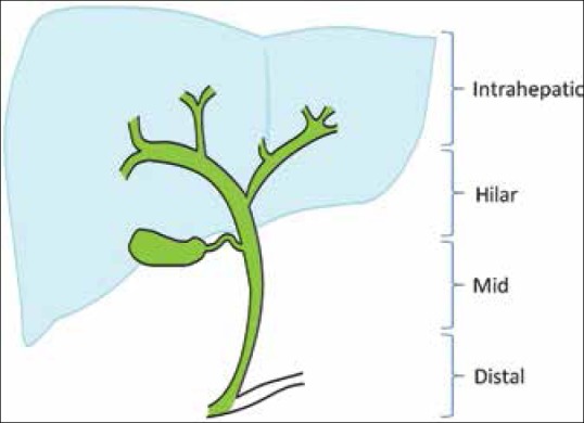
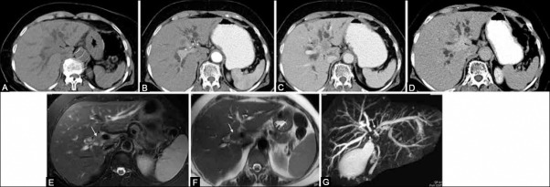
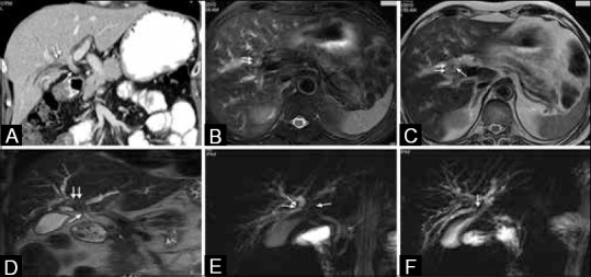
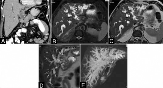
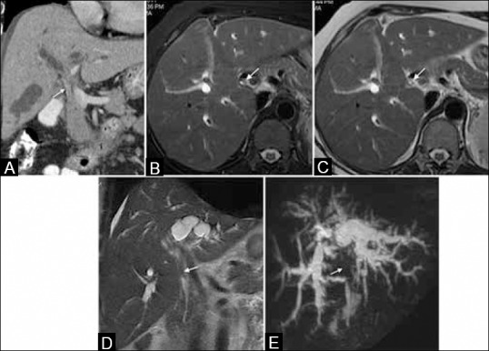
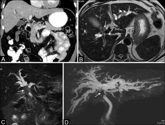
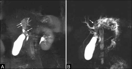
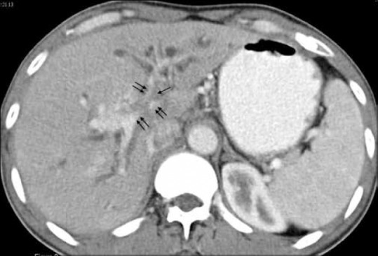
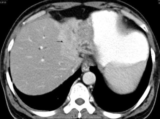
Although hilar cholangiocarcinoma is relatively rare, it can be diagnosed on imaging by identifying its typical pattern. In most cases, the tumor appears to be centered on the right or left hepatic duct with involvement of the ipsilateral portal vein, atrophy of hepatic lobe on that side, and invasion of adjacent liver parenchyma. Multi-detector computed tomography (MDCT) and magnetic resonance cholangiopancreatography (MRCP) are commonly used imaging modalities to assess the longitudinal and horizontal spread of tumor.
尽管肝门部胆管癌相对少见,但通过识别其典型表现,可在影像学上作出诊断。在大多数情况下,肿瘤似乎以左右肝管为中心,累及同侧门静脉,该侧肝叶萎缩,并侵犯相邻肝实质。多排螺旋计算机断层扫描(MDCT)和磁共振胰胆管造影(MRCP)是常用的影像学检查方法,用于评估肿瘤的纵向和横向扩散情况。