Kratz Megan B, Manis Paul B
Department of Otolaryngology/Head and Neck Surgery, University of North Carolina at Chapel Hill Chapel Hill, NC, USA ; The Curriculum in Neurobiology, University of North Carolina Chapel Hill, NC, USA.
Department of Otolaryngology/Head and Neck Surgery, University of North Carolina at Chapel Hill Chapel Hill, NC, USA ; The Curriculum in Neurobiology, University of North Carolina Chapel Hill, NC, USA ; Department of Cell Biology and Physiology, University of North Carolina Chapel Hill, NC, USA.
Front Neural Circuits. 2015 Apr 29;9:17. doi: 10.3389/fncir.2015.00017. eCollection 2015.
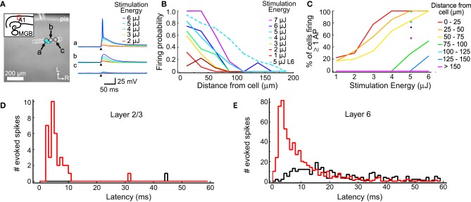
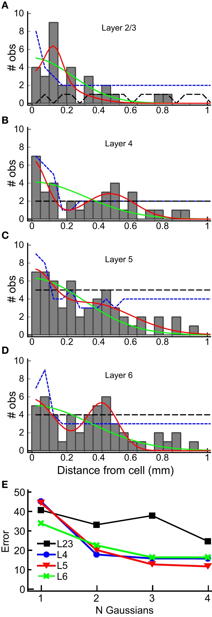
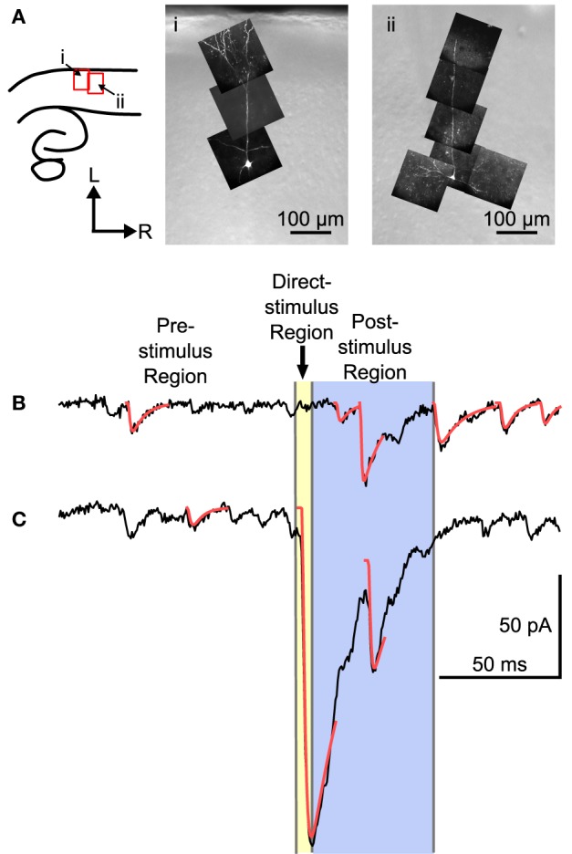
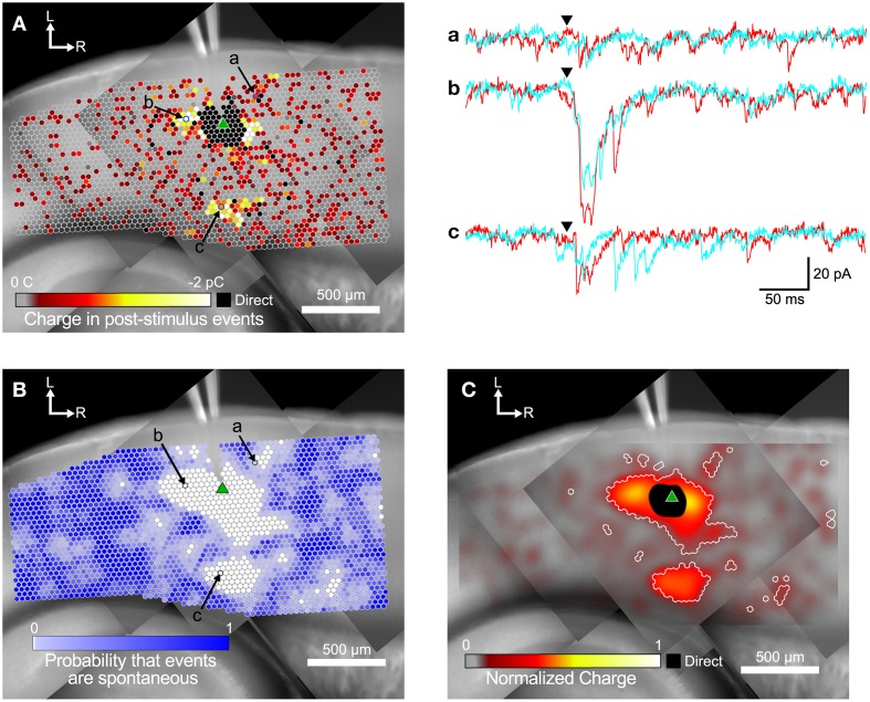
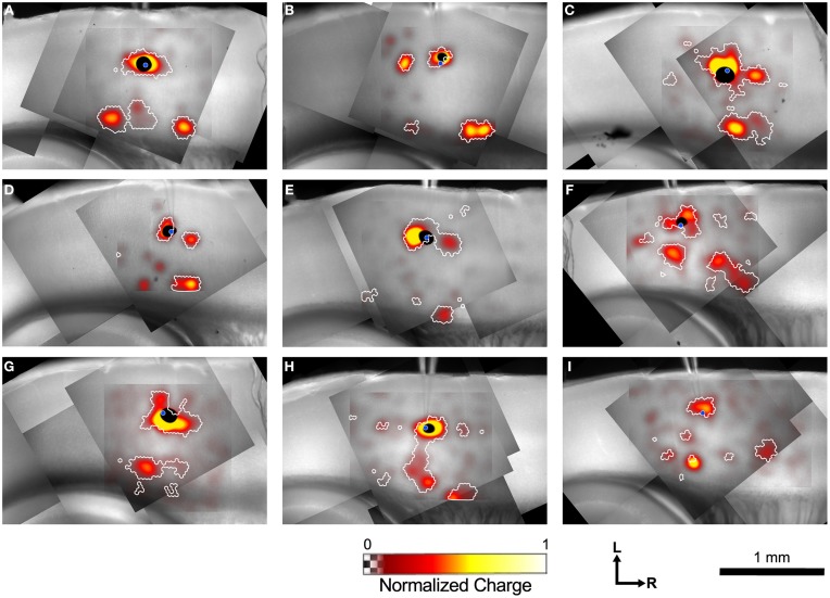
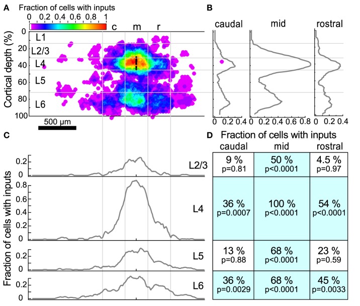
Layer 4 (L4) of primary auditory cortex (A1) receives a tonotopically organized projection from the medial geniculate nucleus of the thalamus. However, individual neurons in A1 respond to a wider range of sound frequencies than would be predicted by their thalamic input, which suggests the existence of cross-frequency intracortical networks. We used laser scanning photostimulation and uncaging of glutamate in brain slices of mouse A1 to characterize the spatial organization of intracortical inputs to L4 neurons. Slices were prepared to include the entire tonotopic extent of A1. We find that L4 neurons receive local vertically organized (columnar) excitation from layers 2 through 6 (L6) and horizontally organized excitation primarily from L4 and L6 neurons in regions centered ~300-500 μm caudal and/or rostral to the cell. Excitatory horizontal synaptic connections from layers 2 and 3 were sparse. The origins of horizontal projections from L4 and L6 correspond to regions in the tonotopic map that are approximately an octave away from the target cell location. Such spatially organized lateral connections may contribute to the detection and processing of auditory objects with specific spectral structures.
初级听觉皮层(A1)的第4层(L4)接收来自丘脑内侧膝状体核的音调拓扑组织投射。然而,A1中的单个神经元对声音频率的响应范围比其丘脑输入所预测的范围更广,这表明存在跨频率的皮质内网络。我们使用激光扫描光刺激和在小鼠A1脑片中释放谷氨酸来表征L4神经元皮质内输入的空间组织。制备的脑片包含A1的整个音调拓扑范围。我们发现,L4神经元从第2层到第6层(L6)接收局部垂直组织(柱状)的兴奋性输入,并且主要从细胞尾侧和/或头侧约300 - 500μm处的区域中的L4和L6神经元接收水平组织的兴奋性输入。来自第2层和第3层的兴奋性水平突触连接稀疏。来自L4和L6的水平投射起源对应于音调拓扑图中与目标细胞位置大约相差一个八度的区域。这种空间组织的侧向连接可能有助于检测和处理具有特定频谱结构的听觉对象。