• 文献检索
  • 文档翻译
  • 深度研究
  • 学术资讯
  • Suppr Zotero 插件Zotero 插件
  • 邀请有礼
  • 套餐&价格
  • 历史记录
应用&插件
Suppr Zotero 插件Zotero 插件浏览器插件Mac 客户端Windows 客户端微信小程序
定价
高级版会员购买积分包购买API积分包
服务
文献检索文档翻译深度研究API 文档MCP 服务
关于我们
关于 Suppr公司介绍联系我们用户协议隐私条款
关注我们

Suppr 超能文献

核心技术专利:CN118964589B侵权必究
粤ICP备2023148730 号-1Suppr @ 2026

文献检索

告别复杂PubMed语法,用中文像聊天一样搜索,搜遍4000万医学文献。AI智能推荐,让科研检索更轻松。

立即免费搜索

文件翻译

保留排版,准确专业,支持PDF/Word/PPT等文件格式,支持 12+语言互译。

免费翻译文档

深度研究

AI帮你快速写综述,25分钟生成高质量综述,智能提取关键信息,辅助科研写作。

立即免费体验

外源性Nkx2.5或GATA-4转染的兔骨髓间充质干细胞与心肌细胞共培养对兔心肌梗死的治疗作用

Exogenous Nkx2.5- or GATA-4-transfected rabbit bone marrow mesenchymal stem cells and myocardial cell co-culture on the treatment of myocardial infarction in rabbits.

作者信息

Li Pu, Zhang Lei

机构信息

Department of Cardiac Surgery, The Third Hospital of Hebei Medical University, Hebei, Shijiazhuang 050017, P.R. China.

Department of Histology and Embryology, Hebei Medical University, Hebei, Shijiazhuang 050017, P.R. China.

出版信息

Mol Med Rep. 2015 Aug;12(2):2607-21. doi: 10.3892/mmr.2015.3775. Epub 2015 May 12.

DOI:10.3892/mmr.2015.3775
PMID:25975979
原文链接:https://pmc.ncbi.nlm.nih.gov/articles/PMC4464300/
Abstract

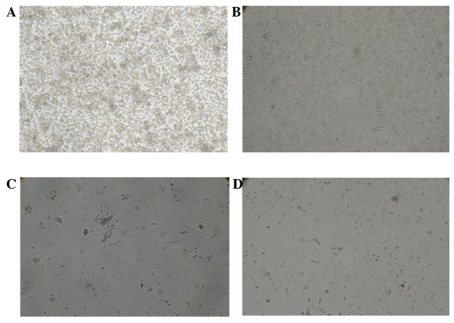
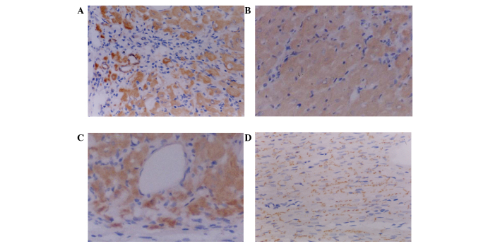
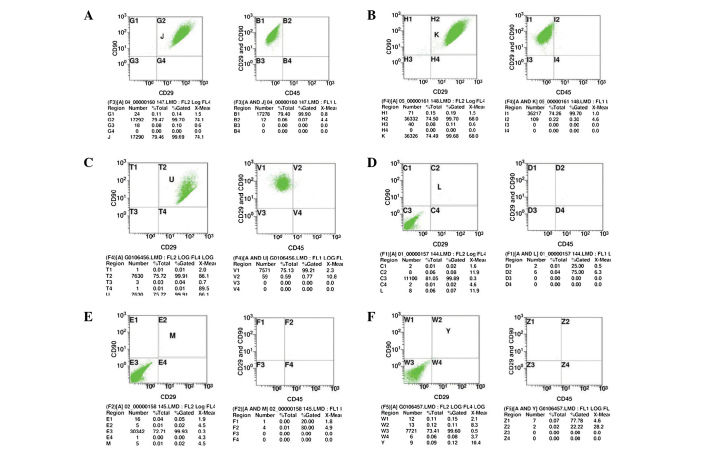
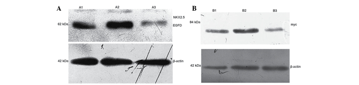
The present study aimed to investigate the effects of Nkx2.5 or GATA-4 transfection with myocardial extracellular environment co-culture on the transformation of bone marrow mesenchymal stem cells (BMSCs) into differentiated cardiomyocytes. Nkx2.5 or GATA-4 were transfected into myocardial extracellular environment co-cultured BMSCs, and then injected into the periphery of infarcted myocardium of a myocardial infarction rabbit model. The effects of these gene transfections and culture on the infarcted myocardium were observed and the results may provide an experimental basis for the efficient myocardial cell differentiation of BMSCs. The present study also suggested that these cells may provide a source and clinical basis for myocardial injury repair via stem cell transplantation. The present study examined whether Nkx2.5 or GATA-4 exogenous gene transfection with myocardial cell extracellular environment co-culture were able to induce the differentiation of BMSCs into cardiac cells. In addition, the effect of these transfected BMSCs on the repair of the myocardium following myocardial infarction was determined using New Zealand rabbit models. The results demonstrated that myocardial cell differentiation was significantly less effective following exogenous gene transfection of Nkx2.5 or GATA-4 alone compared with that of transfection in combination with extracellular environment co-culture. In addition, the results of the present study showed that exogenous gene transfection of Nkx2.5 or GATA-4 into myocardial cell extracellular environment co-cultured BMSCs was able to significantly enhance the ability to repair, mitigating the death of myocardial cells and activation of the myocardium in rabbits with myocardial infarction compared with those of the rabbits transplanted with untreated BMSCs. In conclusion, the exogenous Nkx2.5 and GATA-4 gene transfection into myocardial extracellular environment co-cultured BMSCs induced increased differentiation into myocardial cells compared with that of gene transfection alone. Furthermore, significantly enhanced reparative effects were observed in the myocardium of rabbits following treatment with Nkx2.5-or GATA-4-transfected myocardial cell extracellular environment co-cultured BMSCs compared with those treated with untreated BMSCs.

摘要

本研究旨在探讨在心肌细胞外环境共培养条件下,转染Nkx2.5或GATA-4对骨髓间充质干细胞(BMSCs)向分化心肌细胞转化的影响。将Nkx2.5或GATA-4转染到心肌细胞外环境共培养的BMSCs中,然后注射到心肌梗死兔模型梗死心肌的周边区域。观察这些基因转染和培养对梗死心肌的影响,结果可为BMSCs高效分化为心肌细胞提供实验依据。本研究还表明,这些细胞可能为通过干细胞移植修复心肌损伤提供来源和临床依据。本研究检测了在心肌细胞外环境共培养条件下,转染Nkx2.5或GATA-4外源基因是否能够诱导BMSCs分化为心肌细胞。此外,使用新西兰兔模型确定了这些转染的BMSCs对心肌梗死后心肌修复的作用。结果表明,单独转染Nkx2.5或GATA-4外源基因后,心肌细胞分化效果明显低于与细胞外环境共培养转染的效果。此外,本研究结果显示,与移植未处理BMSCs的兔子相比,将Nkx2.5或GATA-4转染到心肌细胞外环境共培养的BMSCs中,能够显著增强修复能力,减轻心肌梗死兔子心肌细胞的死亡并激活心肌。总之,与单独基因转染相比,将外源Nkx2.5和GATA-4基因转染到心肌细胞外环境共培养的BMSCs中可诱导其向心肌细胞的分化增加。此外,与未处理BMSCs治疗的兔子相比,用Nkx2.5或GATA-4转染的心肌细胞外环境共培养的BMSCs治疗的兔子心肌修复效果显著增强。

https://cdn.ncbi.nlm.nih.gov/pmc/blobs/6ba8/4464300/6ff606817904/MMR-12-02-2607-g11.jpg
https://cdn.ncbi.nlm.nih.gov/pmc/blobs/6ba8/4464300/159b9adadbce/MMR-12-02-2607-g00.jpg
https://cdn.ncbi.nlm.nih.gov/pmc/blobs/6ba8/4464300/c1f7117e485f/MMR-12-02-2607-g01.jpg
https://cdn.ncbi.nlm.nih.gov/pmc/blobs/6ba8/4464300/4e027a99a1a0/MMR-12-02-2607-g02.jpg
https://cdn.ncbi.nlm.nih.gov/pmc/blobs/6ba8/4464300/372c9af031b4/MMR-12-02-2607-g03.jpg
https://cdn.ncbi.nlm.nih.gov/pmc/blobs/6ba8/4464300/5a308c9b9cfb/MMR-12-02-2607-g04.jpg
https://cdn.ncbi.nlm.nih.gov/pmc/blobs/6ba8/4464300/50f42d9071d6/MMR-12-02-2607-g05.jpg
https://cdn.ncbi.nlm.nih.gov/pmc/blobs/6ba8/4464300/20c7cbf41155/MMR-12-02-2607-g06.jpg
https://cdn.ncbi.nlm.nih.gov/pmc/blobs/6ba8/4464300/689003dbd316/MMR-12-02-2607-g07.jpg
https://cdn.ncbi.nlm.nih.gov/pmc/blobs/6ba8/4464300/406abbab855f/MMR-12-02-2607-g08.jpg
https://cdn.ncbi.nlm.nih.gov/pmc/blobs/6ba8/4464300/f15b489368fd/MMR-12-02-2607-g09.jpg
https://cdn.ncbi.nlm.nih.gov/pmc/blobs/6ba8/4464300/45fec12a6562/MMR-12-02-2607-g10.jpg
https://cdn.ncbi.nlm.nih.gov/pmc/blobs/6ba8/4464300/6ff606817904/MMR-12-02-2607-g11.jpg

相似文献

1
Exogenous Nkx2.5- or GATA-4-transfected rabbit bone marrow mesenchymal stem cells and myocardial cell co-culture on the treatment of myocardial infarction in rabbits.外源性Nkx2.5或GATA-4转染的兔骨髓间充质干细胞与心肌细胞共培养对兔心肌梗死的治疗作用
Mol Med Rep. 2015 Aug;12(2):2607-21. doi: 10.3892/mmr.2015.3775. Epub 2015 May 12.
2
GATA-4-expressing mouse bone marrow mesenchymal stem cells improve cardiac function after myocardial infarction via secreted exosomes.GATA-4 表达的鼠骨髓间充质干细胞通过分泌的外泌体改善心肌梗死后的心功能。
Sci Rep. 2018 Jun 13;8(1):9047. doi: 10.1038/s41598-018-27435-9.
3
Hepatocyte growth factor combined with insulin like growth factor-1 improves expression of GATA-4 in mesenchymal stem cells cocultured with cardiomyocytes.肝细胞生长因子联合胰岛素样生长因子-1可改善与心肌细胞共培养的间充质干细胞中GATA-4的表达。
Chin Med J (Engl). 2008 Feb 20;121(4):336-40.
4
Cardiac differentiation is driven by NKX2.5 and GATA4 nuclear translocation in tissue-specific mesenchymal stem cells.心脏分化是由组织特异性间充质干细胞中的NKX2.5和GATA4核转位驱动的。
Stem Cells Dev. 2009 Jul-Aug;18(6):907-18. doi: 10.1089/scd.2008.0292.
5
Overexpressing NKx2.5 increases the differentiation of human umbilical cord drived mesenchymal stem cells into cardiomyocyte-like cells.过表达NKx2.5可增加人脐带间充质干细胞向心肌样细胞的分化。
Biomed Pharmacother. 2016 Mar;78:110-115. doi: 10.1016/j.biopha.2016.01.020. Epub 2016 Jan 24.
6
Enhanced therapeutic effects of mesenchymal stem cells on myocardial infarction by ischemic postconditioning through paracrine mechanisms in rats.缺血后处理通过旁分泌机制增强大鼠骨髓间充质干细胞治疗心肌梗死的疗效。
J Mol Cell Cardiol. 2011 Nov;51(5):839-47. doi: 10.1016/j.yjmcc.2011.06.013. Epub 2011 Jun 29.
7
Overexpression of Csx/Nkx2.5 and GATA-4 enhances the efficacy of mesenchymal stem cell transplantation after myocardial infarction.Csx/Nkx2.5 和 GATA-4 的过表达增强了心肌梗死后间充质干细胞移植的疗效。
Circ J. 2011;75(11):2683-91. doi: 10.1253/circj.cj-11-0238. Epub 2011 Aug 9.
8
[The expression of GATA-4 and Nkx2.5 gene in the transformation of Rattus mesenchymal stem cells into cardiomyocytes].[GATA-4和Nkx2.5基因在大鼠间充质干细胞向心肌细胞转化中的表达]
Sichuan Da Xue Xue Bao Yi Xue Ban. 2008 Nov;39(6):882-5.
9
A brief review: the therapeutic potential of bone marrow mesenchymal stem cells in myocardial infarction.简要综述:骨髓间充质干细胞在心肌梗死中的治疗潜力。
Stem Cell Res Ther. 2017 Nov 2;8(1):242. doi: 10.1186/s13287-017-0697-9.
10
Delayed enrichment for c-kit and inducing cardiac differentiation attenuated protective effects of BMSCs' transplantation in pig model of acute myocardial ischemia.c-kit的延迟富集和诱导心脏分化减弱了骨髓间充质干细胞移植在猪急性心肌缺血模型中的保护作用。
Cardiovasc Ther. 2015 Aug;33(4):184-92. doi: 10.1111/1755-5922.12131.

引用本文的文献

1
The Role of Stem Cells in the Treatment of Cardiovascular Diseases.干细胞在心血管疾病治疗中的作用。
Int J Mol Sci. 2024 Mar 31;25(7):3901. doi: 10.3390/ijms25073901.
2
Mineralocorticoid receptor deficiency improves the therapeutic effects of mesenchymal stem cells for myocardial infarction via enhanced cell survival.醛固酮受体缺乏通过增强细胞存活改善间充质干细胞治疗心肌梗死的疗效。
J Cell Mol Med. 2019 Feb;23(2):1246-1256. doi: 10.1111/jcmm.14026. Epub 2018 Dec 13.
3
Direct Cardiac Reprogramming: A Novel Approach for Heart Regeneration.

本文引用的文献

1
Isolation and cellular properties of mesenchymal cells derived from the decidua of human term placenta.人足月胎盘中蜕膜来源的间充质细胞的分离和细胞特性。
Differentiation. 2011 Sep;82(2):77-88. doi: 10.1016/j.diff.2011.05.010.
2
In vitro Differentiation Potential of Mesenchymal Stem Cells.间充质干细胞的体外分化潜能
Transfus Med Hemother. 2008;35(3):228-238. doi: 10.1159/000124281. Epub 2008 May 8.
3
Comparative analysis of telomere length, telomerase and reverse transcriptase activity in human dental stem cells.人牙髓干细胞中端粒长度、端粒酶和逆转录酶活性的比较分析。
直接心脏重编程:心脏再生的新方法。
Int J Mol Sci. 2018 Sep 5;19(9):2629. doi: 10.3390/ijms19092629.
4
Reactive Oxygen Species, Superoxide Dimutases, and PTEN-p53-AKT-MDM2 Signaling Loop Network in Mesenchymal Stem/Stromal Cells Regulation.间充质干/基质细胞调控中的活性氧、超氧化物歧化酶以及PTEN-p53-AKT-MDM2信号环路网络
Cells. 2018 May 1;7(5):36. doi: 10.3390/cells7050036.
5
Mesenchymal stem cells in cardiac regeneration: a detailed progress report of the last 6 years (2010-2015).心脏再生中的间充质干细胞:过去6年(2010 - 2015年)的详细进展报告
Stem Cell Res Ther. 2016 Jun 4;7(1):82. doi: 10.1186/s13287-016-0341-0.
6
Direct intercellular communications dominate the interaction between adipose-derived MSCs and myofibroblasts against cardiac fibrosis.直接的细胞间通讯在脂肪来源的间充质干细胞与肌成纤维细胞对抗心脏纤维化的相互作用中起主导作用。
Protein Cell. 2015 Oct;6(10):735-45. doi: 10.1007/s13238-015-0196-7. Epub 2015 Aug 14.
Cell Transplant. 2011;20(11-12):1693-705. doi: 10.3727/096368911X565001. Epub 2011 Mar 8.
4
Impact of anti-apoptotic and anti-oxidative effects of bone marrow mesenchymal stem cells with transient overexpression of heme oxygenase-1 on myocardial ischemia.骨髓间充质干细胞过表达血红素氧合酶-1的抗细胞凋亡和抗氧化作用对心肌缺血的影响。
Am J Physiol Heart Circ Physiol. 2010 May;298(5):H1320-9. doi: 10.1152/ajpheart.01330.2008. Epub 2010 Feb 12.
5
In vitro and in vivo evaluation of osteogenesis of human umbilical cord blood-derived mesenchymal stem cells on partially demineralized bone matrix.人脐带血间充质干细胞在部分脱矿骨基质上的体外和体内成骨评价。
Tissue Eng Part A. 2010 Mar;16(3):971-82. doi: 10.1089/ten.TEA.2009.0516.
6
In vitro and in vivo osteogenesis of porcine skin-derived mesenchymal stem cell-like cells with a demineralized bone and fibrin glue scaffold.脱矿骨与纤维蛋白胶支架诱导猪皮肤来源间充质干细胞样细胞的体外与体内成骨。
Tissue Eng Part A. 2010 Mar;16(3):815-27. doi: 10.1089/ten.TEA.2009.0439.
7
Fresh fields and pathways new: recent genetic insights into cardiac malformation.新的领域与途径:心脏畸形的最新遗传学见解
Heart. 2009 Mar;95(6):442-7. doi: 10.1136/hrt.2006.105130.
8
Cooperative interaction of Nkx2.5 and Mef2c transcription factors during heart development.心脏发育过程中Nkx2.5和Mef2c转录因子的协同相互作用。
Dev Dyn. 2008 Dec;237(12):3809-19. doi: 10.1002/dvdy.21803.
9
Effect of acute and chronic simvastatin treatment on post-ischemic contractile dysfunction in isolated rat heart.急性和慢性辛伐他汀治疗对离体大鼠心脏缺血后收缩功能障碍的影响。
Physiol Res. 2008;57(5):793-796. doi: 10.33549/physiolres.931559.
10
Evaluation of partially demineralized osteoporotic cancellous bone matrix combined with human bone marrow stromal cells for tissue engineering: an in vitro and in vivo study.部分脱矿质骨质疏松性松质骨基质与人骨髓基质细胞联合用于组织工程的评估:一项体内外研究
Calcif Tissue Int. 2008 Sep;83(3):176-85. doi: 10.1007/s00223-008-9159-9. Epub 2008 Aug 15.