Zhao Yu, Deng Bin, Li Yichong, Zhou Lihua, Yang Lei, Gou Xingchun, Wang Qiang, Chen Guozhong, Xu Hao, Xu Lixian
State Key Laboratory of Military Stomatology, Department of Anesthesiology, School of Stomatology, The Fourth Military Medical University, Xi'an, 710032, China.
Department of Anesthesiology, Binghua Hospital, Haerbin, 150080, China.
Cell Mol Neurobiol. 2015 Nov;35(8):1093-103. doi: 10.1007/s10571-015-0203-9. Epub 2015 May 15.
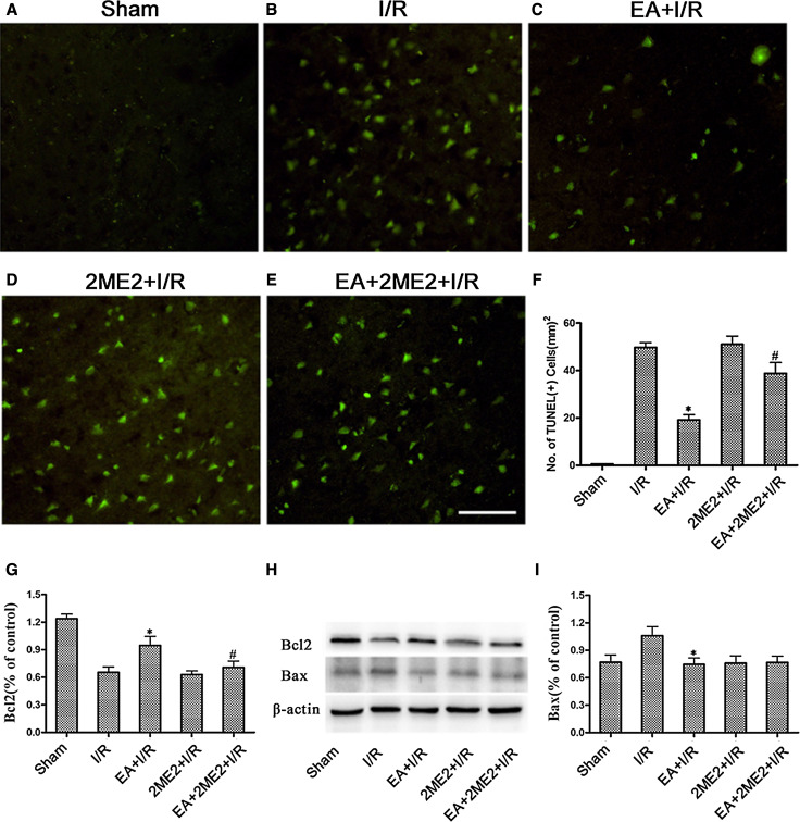
We have reported electroacupuncture (EA) pretreatment induced the tolerance against focal cerebral ischemia through activation of canonical Notch pathway. However, the underlying mechanisms have not been fully understood. Evidences suggest that up-regulation of hypoxia inducible factor-1α (HIF-1α) contributes to neuroprotection against ischemia which could interact with Notch signaling pathway in this process. Therefore, the current study is to test that up-regulation of HIF-1α associated with Notch pathway contributes to the neuroprotection of EA pretreatment. Sprague-Dawley rats were treated with EA at the acupoint "Baihui (GV 20)" 30 min per day for successive 5 days before MCAO. HIF-1α levels were measured before and after reperfusion. Then, HIF-1α antagonist 2ME2 and γ-secretase inhibitor MW167 were used. Neurologic deficit scores, infarction volumes, neuronal apoptosis, and Bcl2/Bax were evaluated. HIF-1α and Notch1 intracellular domain (NICD) were assessed. The results showed EA pretreatment enhanced the neuronal expression of HIF-1α, reduced infarct volume, improved neurological outcome, inhibited neuronal apoptosis, up-regulated expression of Bcl-2, and down-regulated expression of Bax after reperfusion in the penumbra, while the beneficial effects were attenuated by 2ME2. Furthermore, intraventricular injection with MW167 efficiently suppressed both up-regulation of NICD and HIF-1α after reperfusion. However, administration with 2ME2 could only decrease the expression of HIF-1α in the penumbra. In conclusion, EA pretreatment exerts neuroprotection against ischemic injury through Notch pathway-mediated up-regulation of HIF-1α.
我们曾报道电针预处理通过激活经典Notch信号通路诱导对局灶性脑缺血的耐受性。然而,其潜在机制尚未完全明确。有证据表明,缺氧诱导因子-1α(HIF-1α)的上调有助于对缺血的神经保护,在此过程中其可能与Notch信号通路相互作用。因此,本研究旨在验证与Notch通路相关的HIF-1α上调有助于电针预处理的神经保护作用。在大脑中动脉闭塞(MCAO)前,将Sprague-Dawley大鼠每天在“百会(GV 20)”穴位进行30分钟电针治疗,连续5天。在再灌注前后测量HIF-1α水平。然后,使用HIF-1α拮抗剂2ME2和γ-分泌酶抑制剂MW167。评估神经功能缺损评分、梗死体积、神经元凋亡以及Bcl2/Bax比值。检测HIF-1α和Notch1细胞内结构域(NICD)。结果显示,电针预处理增强了半暗带区再灌注后神经元HIF-1α的表达,减小了梗死体积,改善了神经功能结局,抑制了神经元凋亡,上调了Bcl-2的表达,下调了Bax的表达,而2ME2减弱了这些有益作用。此外,脑室内注射MW167有效抑制了再灌注后NICD和HIF-1α的上调。然而,给予2ME2仅能降低半暗带区HIF-1α 的表达。总之,电针预处理通过Notch通路介导的HIF-1α上调发挥对缺血性损伤的神经保护作用。