Carvalho Bettina, Hamerschmidt Rogerio, Telles Jose Ederaldo, Richter Nicole
Department of ENT, IPO, Curitiba, PR, Brazil.
Department of Pathology, HC/UFPR, Curitiba, PR, Brazil.
Int Arch Otorhinolaryngol. 2015 Jan;19(1):1-4. doi: 10.1055/s-0034-1382096. Epub 2014 Jul 18.
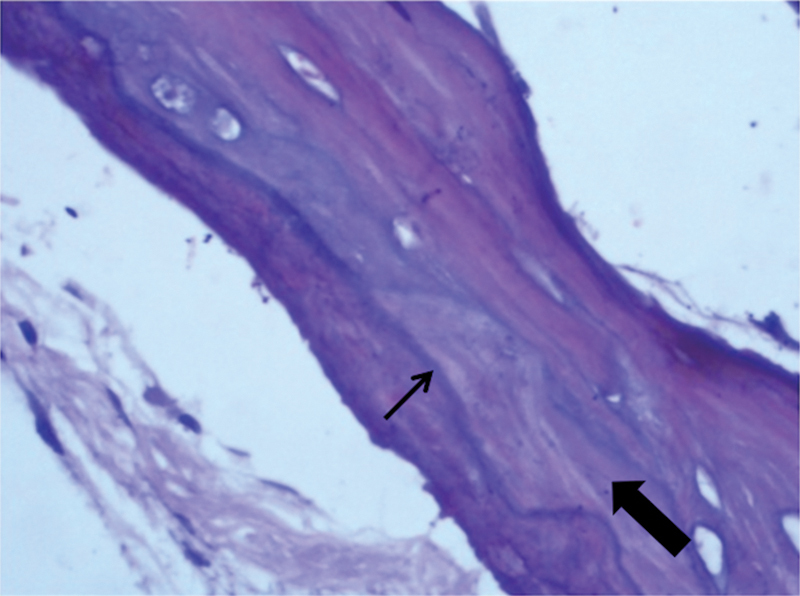
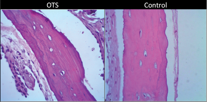
Introduction Otosclerosis is a disease that causes bone resorption and deposition in the auditory structures, leading to deafness. Many studies have evaluated the histopathology of the stapes footplate in this disease (osteoblasts, osteoclasts, vascular proliferation, fibroblasts, and histiocytes), but we found no studies in the literature involving the histology of the superstructure of the stapes. Objectives To perform an analysis under optical microscopy of histopathologic findings of the superstructure of the stapes from patients with otosclerosis. Methods A contemporary cross-sectional cohort study of pathology analysis of superstructures of the stapes of patients with otosclerosis. Results Fifteen superstructures of stapes in patients with otosclerosis operated in our service and four stapes of cadavers used for dissection (controls) were evaluated. No areas of bone resorption or deposition or presence of osteoclasts and osteoblasts in the superstructure of the stapes were found. However, we found in the more distal portions of the crura areas with prominent cementitious lines and woven bone, which was different than the mature trabecular bone found in the head of the stapes or in the controls. Conclusion There were histologic changes in the superstructure of the stapes in patients with otosclerosis operated in our service.
引言 耳硬化症是一种导致听觉结构中骨质吸收和沉积,进而引发耳聋的疾病。许多研究评估了该疾病中镫骨足板的组织病理学(成骨细胞、破骨细胞、血管增生、成纤维细胞和组织细胞),但我们在文献中未发现涉及镫骨上部结构组织学的研究。目的 对耳硬化症患者镫骨上部结构的组织病理学发现进行光学显微镜分析。方法 对耳硬化症患者镫骨上部结构进行病理学分析的当代横断面队列研究。结果 评估了在我们科室接受手术的耳硬化症患者的15个镫骨上部结构以及4个用于解剖的尸体镫骨(对照)。在镫骨上部结构中未发现骨质吸收或沉积区域,也未发现破骨细胞和成骨细胞。然而,我们在镫骨脚的更远端部分发现了有明显黏合线和编织骨的区域,这与镫骨头或对照中发现的成熟小梁骨不同。结论 在我们科室接受手术的耳硬化症患者的镫骨上部结构存在组织学变化。