• 文献检索
  • 文档翻译
  • 深度研究
  • 学术资讯
  • Suppr Zotero 插件Zotero 插件
  • 邀请有礼
  • 套餐&价格
  • 历史记录
应用&插件
Suppr Zotero 插件Zotero 插件浏览器插件Mac 客户端Windows 客户端微信小程序
定价
高级版会员购买积分包购买API积分包
服务
文献检索文档翻译深度研究API 文档MCP 服务
关于我们
关于 Suppr公司介绍联系我们用户协议隐私条款
关注我们

Suppr 超能文献

核心技术专利:CN118964589B侵权必究
粤ICP备2023148730 号-1Suppr @ 2026

文献检索

告别复杂PubMed语法,用中文像聊天一样搜索,搜遍4000万医学文献。AI智能推荐,让科研检索更轻松。

立即免费搜索

文件翻译

保留排版,准确专业,支持PDF/Word/PPT等文件格式,支持 12+语言互译。

免费翻译文档

深度研究

AI帮你快速写综述,25分钟生成高质量综述,智能提取关键信息,辅助科研写作。

立即免费体验

一种用于在扩散加权磁共振成像上对疑似甲状腺肿瘤进行术前分层的新型纹理分析工具的多机构验证

Multi-institutional validation of a novel textural analysis tool for preoperative stratification of suspected thyroid tumors on diffusion-weighted MRI.

作者信息

Brown Anna M, Nagala Sidhartha, McLean Mary A, Lu Yonggang, Scoffings Daniel, Apte Aditya, Gonen Mithat, Stambuk Hilda E, Shaha Ashok R, Tuttle R Michael, Deasy Joseph O, Priest Andrew N, Jani Piyush, Shukla-Dave Amita, Griffiths John

机构信息

Cancer Research UK Cambridge Institute, University of Cambridge, Li Ka Shing Centre, Robinson Way, Cambridge, United Kingdom.

Duke University School of Medicine, Durham, North Carolina, USA.

出版信息

Magn Reson Med. 2016 Apr;75(4):1708-16. doi: 10.1002/mrm.25743. Epub 2015 May 20.

DOI:10.1002/mrm.25743
PMID:25995019
原文链接:https://pmc.ncbi.nlm.nih.gov/articles/PMC4654719/
Abstract

PURPOSE

Ultrasound-guided fine needle aspirate cytology fails to diagnose many malignant thyroid nodules; consequently, patients may undergo diagnostic lobectomy. This study assessed whether textural analysis (TA) could noninvasively stratify thyroid nodules accurately using diffusion-weighted MRI (DW-MRI).

METHODS

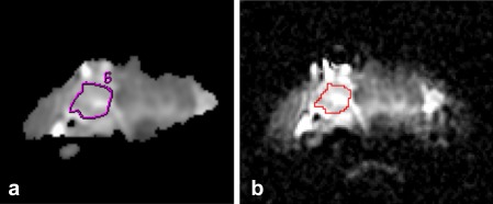
This multi-institutional study examined 3T DW-MRI images obtained with spin echo echo planar imaging sequences. The training data set included 26 patients from Cambridge, United Kingdom, and the test data set included 18 thyroid cancer patients from Memorial Sloan Kettering Cancer Center (New York, New York, USA). Apparent diffusion coefficients (ADCs) were compared over regions of interest (ROIs) defined on thyroid nodules. TA, linear discriminant analysis (LDA), and feature reduction were performed using the 21 MaZda-generated texture parameters that best distinguished benign and malignant ROIs.

RESULTS

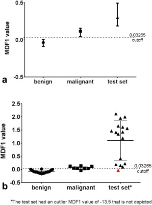
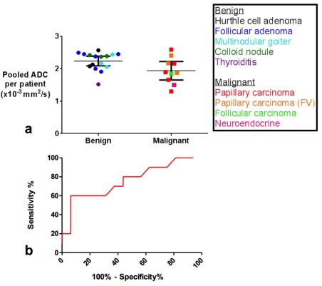
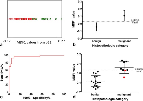
Training data set mean ADC values were significantly different for benign and malignant nodules (P = 0.02) with a sensitivity and specificity of 70% and 63%, respectively, and a receiver operator characteristic (ROC) area under the curve (AUC) of 0.73. The LDA model of the top 21 textural features correctly classified 89/94 DW-MRI ROIs with 92% sensitivity, 96% specificity, and an AUC of 0.97. This algorithm correctly classified 16/18 (89%) patients in the independently obtained test set of thyroid DW-MRI scans.

CONCLUSION

TA classifies thyroid nodules with high sensitivity and specificity on multi-institutional DW-MRI data sets. This method requires further validation in a larger prospective study. Magnetic Resonance in Medicine published by Wiley Periodicals, Inc. on behalf of International Society for Magnetic Resonance.

摘要

目的

超声引导下细针穿刺抽吸细胞学检查无法诊断许多恶性甲状腺结节;因此,患者可能需要接受诊断性肺叶切除术。本研究评估了纹理分析(TA)能否使用扩散加权磁共振成像(DW-MRI)对甲状腺结节进行准确的无创分层。

方法

这项多机构研究检查了通过自旋回波平面回波成像序列获得的3T DW-MRI图像。训练数据集包括来自英国剑桥的26名患者,测试数据集包括来自美国纽约纪念斯隆凯特琳癌症中心的18名甲状腺癌患者。比较了在甲状腺结节上定义的感兴趣区域(ROI)的表观扩散系数(ADC)。使用21个由MaZda生成的能最佳区分良性和恶性ROI的纹理参数进行TA、线性判别分析(LDA)和特征约简。

结果

训练数据集中良性和恶性结节的平均ADC值有显著差异(P = 0.02),敏感性和特异性分别为70%和63%,曲线下面积(AUC)为0.73。前21个纹理特征的LDA模型正确分类了94个DW-MRI ROI中的89个,敏感性为92%,特异性为96%,AUC为0.97。该算法在独立获得的甲状腺DW-MRI扫描测试集中正确分类了16/18(89%)的患者。

结论

TA在多机构DW-MRI数据集上对甲状腺结节进行分类具有高敏感性和特异性。该方法需要在更大规模的前瞻性研究中进一步验证。《医学磁共振》由威利期刊公司代表国际磁共振学会出版。

https://cdn.ncbi.nlm.nih.gov/pmc/blobs/c4c5/4791090/91e932eb5789/MRM-75-1708-g004.jpg
https://cdn.ncbi.nlm.nih.gov/pmc/blobs/c4c5/4791090/77b57560bb21/MRM-75-1708-g001.jpg
https://cdn.ncbi.nlm.nih.gov/pmc/blobs/c4c5/4791090/813f3a571558/MRM-75-1708-g002.jpg
https://cdn.ncbi.nlm.nih.gov/pmc/blobs/c4c5/4791090/abad4ac6055d/MRM-75-1708-g003.jpg
https://cdn.ncbi.nlm.nih.gov/pmc/blobs/c4c5/4791090/91e932eb5789/MRM-75-1708-g004.jpg

相似文献

1
Multi-institutional validation of a novel textural analysis tool for preoperative stratification of suspected thyroid tumors on diffusion-weighted MRI.一种用于在扩散加权磁共振成像上对疑似甲状腺肿瘤进行术前分层的新型纹理分析工具的多机构验证
Magn Reson Med. 2016 Apr;75(4):1708-16. doi: 10.1002/mrm.25743. Epub 2015 May 20.
2
Differentiation between malignant and benign thyroid nodules and stratification of papillary thyroid cancer with aggressive histological features: Whole-lesion diffusion-weighted imaging histogram analysis.甲状腺良恶性结节的鉴别及具有侵袭性组织学特征的甲状腺乳头状癌的分层:全病灶扩散加权成像直方图分析
J Magn Reson Imaging. 2016 Dec;44(6):1546-1555. doi: 10.1002/jmri.25290. Epub 2016 Apr 19.
3
Role of apparent diffusion coefficient values and diffusion-weighted magnetic resonance imaging in differentiation between benign and malignant thyroid nodules.表观扩散系数值及弥散加权磁共振成像在甲状腺良恶性结节鉴别诊断中的作用。
Clin Imaging. 2012 Jan-Feb;36(1):1-7. doi: 10.1016/j.clinimag.2011.04.001.
4
Diagnostic efficacy of multiple MRI parameters in differentiating benign vs. malignant thyroid nodules.多种MRI参数在鉴别甲状腺良恶性结节中的诊断效能
BMC Med Imaging. 2018 Dec 3;18(1):50. doi: 10.1186/s12880-018-0294-0.
5
Quantitative evaluation of benign and malignant vertebral fractures with diffusion-weighted MRI: what is the optimum combination of b values for ADC-based lesion differentiation with the single-shot turbo spin-echo sequence?扩散加权 MRI 对良恶性椎体骨折的定量评估:单次激发快速自旋回波序列基于 ADC 值的病变鉴别最佳 b 值组合是什么?
AJR Am J Roentgenol. 2014 Sep;203(3):582-8. doi: 10.2214/AJR.13.11632.
6
[Value of radiomics models based on MRI diffusion weighted imaging and apparent diffusion coefficient in differentiating benign and malignant thyroid nodules].基于MRI扩散加权成像和表观扩散系数的影像组学模型在鉴别甲状腺良恶性结节中的价值
Zhonghua Yi Xue Za Zhi. 2023 Nov 7;103(41):3279-3286. doi: 10.3760/cma.j.cn112137-20230913-00453.
7
Differential diagnosis of benign and malignant breast masses using diffusion-weighted magnetic resonance imaging.使用扩散加权磁共振成像对乳腺良恶性肿块进行鉴别诊断。
World J Surg Oncol. 2015 Feb 7;13:32. doi: 10.1186/s12957-014-0431-3.
8
Diffusion-weighted MR Imaging and ADC Mapping in Differentiating Benign from Malignant Thyroid Nodules.扩散加权磁共振成像及表观扩散系数图在鉴别甲状腺良恶性结节中的应用
J Coll Physicians Surg Pak. 2015 Nov;25(11):785-8.
9
Diffusion-weighted MR imaging of thyroid nodules.甲状腺结节的扩散加权磁共振成像。
Neuroradiology. 2009 Mar;51(3):193-8. doi: 10.1007/s00234-008-0494-3. Epub 2009 Jan 23.
10
Utility of high b-value (2000 sec/mm2) DWI with RESOLVE in differentiating papillary thyroid carcinomas and papillary thyroid microcarcinomas from benign thyroid nodules.高 b 值(2000 sec/mm 2 )弥散加权成像联合 RESOLVE 技术鉴别甲状腺乳头状癌及微小癌与甲状腺良性结节的价值。
PLoS One. 2018 Jul 18;13(7):e0200270. doi: 10.1371/journal.pone.0200270. eCollection 2018.

引用本文的文献

1
Magnetic resonance imaging-based radiomics signature for predicting preoperative staging of esophageal cancer.基于磁共振成像的放射组学特征用于预测食管癌术前分期
World J Radiol. 2025 Aug 28;17(8):110307. doi: 10.4329/wjr.v17.i8.110307.
2
Diagnostic Accuracy of Diffusion-Weighted MRI for Differentiating Benign and Malignant Thyroid Nodules: Systematic Review and Meta-Analysis.扩散加权磁共振成像鉴别甲状腺良恶性结节的诊断准确性:系统评价与Meta分析
Cancers (Basel). 2025 Aug 18;17(16):2677. doi: 10.3390/cancers17162677.
3
Impact of artificial intelligence-based and traditional image preprocessing and resampling on MRI-based radiomics for classification of papillary thyroid carcinoma.

本文引用的文献

1
3T diffusion-weighted MRI of the thyroid gland with reduced distortion: preliminary results.3T 弥散加权磁共振成像对甲状腺的低失真:初步结果。
Br J Radiol. 2013 Aug;86(1028):20130022. doi: 10.1259/bjr.20130022. Epub 2013 Jun 14.
2
The efficacy of apparent diffusion coefficient value calculation in differentiation between malignant and benign thyroid nodules.表观弥散系数值计算在鉴别甲状腺良恶性结节中的作用。
Clin Imaging. 2012 Jul-Aug;36(4):316-22. doi: 10.1016/j.clinimag.2011.10.006. Epub 2012 Jun 8.
3
Thyroid nodule recognition based on feature selection and pixel classification methods.
基于人工智能和传统的图像预处理及重采样对基于MRI的甲状腺乳头状癌分类的影像组学的影响。
BJR Artif Intell. 2025 Apr 15;2(1):ubaf006. doi: 10.1093/bjrai/ubaf006. eCollection 2025 Jan.
4
Radiomics Applications in Head and Neck Tumor Imaging: A Narrative Review.放射组学在头颈部肿瘤成像中的应用:一篇综述
Cancers (Basel). 2023 Feb 12;15(4):1174. doi: 10.3390/cancers15041174.
5
Radiomic analysis based on multi-phase magnetic resonance imaging to predict preoperatively microvascular invasion in hepatocellular carcinoma.基于多期磁共振成像的放射组学分析预测肝细胞癌术前微血管侵犯。
World J Gastroenterol. 2022 Jun 28;28(24):2733-2747. doi: 10.3748/wjg.v28.i24.2733.
6
Refining Tumor Treatment in Sinonasal Cancer Using Delta Radiomics of Multi-Parametric MRI after the First Cycle of Induction Chemotherapy.在诱导化疗的第一个周期后,利用多参数MRI的增量放射组学优化鼻窦癌的肿瘤治疗。
J Imaging. 2022 Feb 15;8(2):46. doi: 10.3390/jimaging8020046.
7
Magnetic Fields and Cancer: Epidemiology, Cellular Biology, and Theranostics.磁场与癌症:流行病学、细胞生物学与治疗学。
Int J Mol Sci. 2022 Jan 25;23(3):1339. doi: 10.3390/ijms23031339.
8
Machine Learning: Applications and Advanced Progresses of Radiomics in Endocrine Neoplasms.机器学习:放射组学在内分泌肿瘤中的应用与最新进展
J Oncol. 2021 Oct 11;2021:8615450. doi: 10.1155/2021/8615450. eCollection 2021.
9
Establishing ADC-Based Histogram and Texture Features for Early Treatment-Induced Changes in Head and Neck Squamous Cell Carcinoma.基于表观扩散系数(ADC)建立直方图和纹理特征以检测头颈部鳞状细胞癌早期治疗引起的变化
Front Oncol. 2021 Sep 2;11:708398. doi: 10.3389/fonc.2021.708398. eCollection 2021.
10
Radiomics feature stability of open-source software evaluated on apparent diffusion coefficient maps in head and neck cancer.头颈部癌症表观扩散系数图的开源软件放射组学特征稳定性评估。
Sci Rep. 2021 Sep 3;11(1):17633. doi: 10.1038/s41598-021-96600-4.
基于特征选择和像素分类方法的甲状腺结节识别。
J Digit Imaging. 2013 Feb;26(1):119-28. doi: 10.1007/s10278-012-9475-5.
4
Role of apparent diffusion coefficient values and diffusion-weighted magnetic resonance imaging in differentiation between benign and malignant thyroid nodules.表观扩散系数值及弥散加权磁共振成像在甲状腺良恶性结节鉴别诊断中的作用。
Clin Imaging. 2012 Jan-Feb;36(1):1-7. doi: 10.1016/j.clinimag.2011.04.001.
5
Can dynamic contrast-enhanced magnetic resonance imaging combined with texture analysis differentiate malignant glioneuronal tumors from other glioblastoma?动态对比增强磁共振成像联合纹理分析能否将恶性神经胶质神经元肿瘤与其他胶质母细胞瘤区分开来?
Neurol Res Int. 2012;2012:195176. doi: 10.1155/2012/195176. Epub 2011 Dec 1.
6
Quantitative diffusion-weighted magnetic resonance imaging as a powerful adjunct to fine needle aspiration cytology for assessment of thyroid nodules.定量扩散加权磁共振成像作为细针穿刺细胞学检查的有力辅助手段,用于评估甲状腺结节。
Am J Otolaryngol. 2012 Jul-Aug;33(4):408-16. doi: 10.1016/j.amjoto.2011.10.013. Epub 2011 Dec 6.
7
Targeted single-shot methods for diffusion-weighted imaging in the kidneys.肾脏弥散加权成像的靶向单次激发方法。
J Magn Reson Imaging. 2011 Jun;33(6):1517-25. doi: 10.1002/jmri.22556.
8
Cystic change in thyroid nodules: a confounding factor for real-time qualitative thyroid ultrasound elastography.甲状腺结节的囊性变:实时定性甲状腺超声弹性成像的混杂因素。
Clin Radiol. 2011 Sep;66(9):799-807. doi: 10.1016/j.crad.2011.03.011. Epub 2011 Apr 29.
9
Classification of the thyroid nodules based on characteristic sonographic textural feature and correlated histopathology using hierarchical support vector machines.基于特征性超声纹理特征和分层支持向量机相关组织病理学对甲状腺结节进行分类。
Ultrasound Med Biol. 2010 Dec;36(12):2018-26. doi: 10.1016/j.ultrasmedbio.2010.08.019.
10
Texture-based classification of focal liver lesions on MRI at 3.0 Tesla: a feasibility study in cysts and hemangiomas.基于纹理的 3.0T MRI 肝脏局灶性病变分类:囊肿和血管瘤的可行性研究。
J Magn Reson Imaging. 2010 Aug;32(2):352-9. doi: 10.1002/jmri.22268.