Lim Seong Hoon, Hong Bo Young, Oh Jee Hae, Lee Jong In
Department of Rehabilitation Medicine, St. Vincent's Hospital, College of Medicine, The Catholic University of Korea, Republic of Korea.
Department of Rehabilitation Medicine, Seoul St. Mary's Hospital, College of Medicine, The Catholic University of Korea, Republic of Korea.
J Phys Ther Sci. 2015 Apr;27(4):1261-5. doi: 10.1589/jpts.27.1261. Epub 2015 Apr 30.
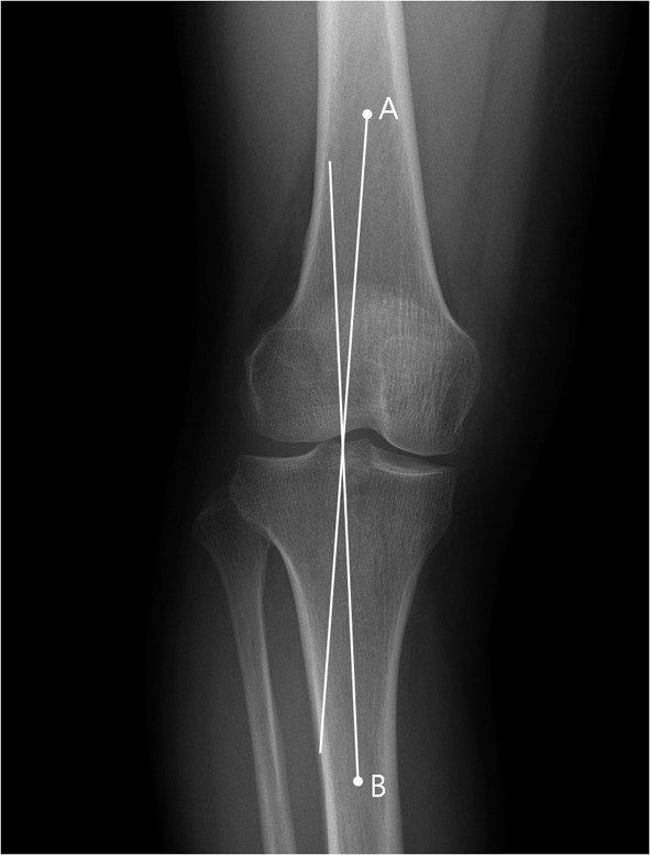
[Purpose] We evaluated the relationship between knee alignment and the electromyographic (EMG) activity of the vastus medialis (VM) to the vastus lateralis (VL) muscles in patients with knee osteoarthritis (OA) in a cross-sectional study. [Subjects and Methods] Forty subjects with knee OA were assessed by anatomic radiographic knee alignment and the VM/VL ratio was calculated. Surface EMG from both the VM and VL muscles were evaluated during maximal isometric contraction at 60° knee flexion. Simultaneously, peak quadriceps torque was assessed using an isokinetic dynamometer. Subjects were categorized into low, moderate, and high varus groups according to knee malalignment. The peak quadriceps torque and VM/VL ratio across groups, and their relationships with varus malalignment were analyzed. [Results] All subjects had medial compartment OA and the VM/VL ratio of all subjects was 1.31 ± 0.28 (mean ± SD). There were no significant differences in the peak quadriceps torque or VM/VL ratios across the groups nor were there any significant relationships with varus malalignment. [Conclusion] The VM/VL ratio and peak quadriceps torque were not associated with the severity of knee varus malalignment.
[目的] 在一项横断面研究中,我们评估了膝关节骨关节炎(OA)患者的膝关节对线与股内侧肌(VM)和股外侧肌(VL)肌电图(EMG)活动之间的关系。[对象与方法] 对40例膝关节OA患者进行解剖学X线膝关节对线评估,并计算VM/VL比值。在膝关节屈曲60°时进行最大等长收缩期间,评估VM和VL肌肉的表面肌电图。同时,使用等速测力计评估股四头肌峰值扭矩。根据膝关节对线不良情况,将受试者分为轻度、中度和重度内翻组。分析各组间的股四头肌峰值扭矩和VM/VL比值,以及它们与内翻对线不良的关系。[结果] 所有受试者均患有内侧间室OA,所有受试者的VM/VL比值为1.31±0.28(平均值±标准差)。各组间的股四头肌峰值扭矩或VM/VL比值无显著差异,与内翻对线不良也无显著关系。[结论] VM/VL比值和股四头肌峰值扭矩与膝关节内翻对线不良的严重程度无关。