Lewek Michael D, Rudolph Katherine S, Snyder-Mackler Lynn
Department of Physical Therapy and Biomechanics and Movement Science Program, University of Delaware, Newark, DE 19716, USA.
Osteoarthritis Cartilage. 2004 Sep;12(9):745-51. doi: 10.1016/j.joca.2004.05.005.
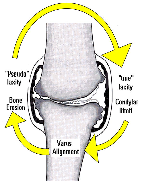
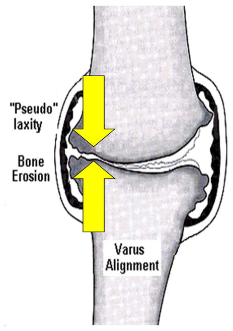
Patients with medial compartment knee osteoarthritis (OA) adopt an abnormal gait pattern, and often develop frontal plane laxity at the knee. The purpose of this study was to quantify the extent of frontal plane knee joint laxity in patients with medial knee OA and genu varum and to assess the effect of joint laxity on knee joint kinetics, kinematics and muscle activity during gait.
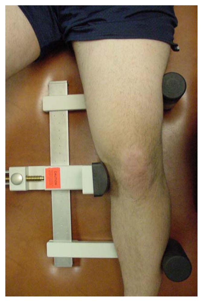
Twelve subjects with genu varum and medial compartment knee osteoarthritis (OA group) and 12 age-matched uninjured subjects underwent stress radiography to determine the presence and magnitude of frontal plane laxity. All subjects also went through gait analysis with surface electromyography of the medial and lateral quadriceps, hamstrings, and gastrocnemius to calculate knee joint kinematics and kinetics and co-contraction levels during gait.
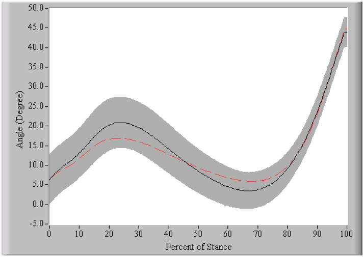
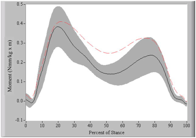
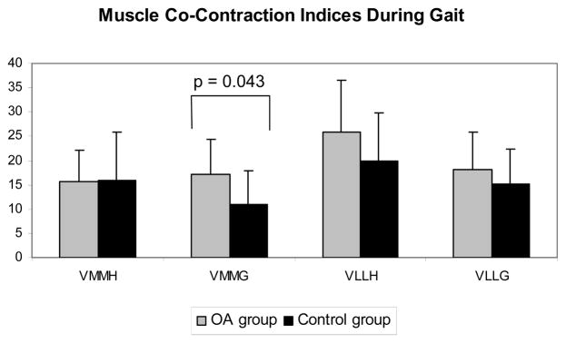
The OA group showed significantly greater knee instability (P = 0.002), medial joint laxity (P = 0.001), greater medial quadriceps-medial gastrocnemius (VMMG) co-contraction (P = 0.043), and greater knee adduction moments (P = 0.019) than the control group. Medial joint laxity contributed significantly to the variance in both VMMG and the knee adduction moment during early stance.
The presence of medial laxity in patients with knee OA is likely contributing to the altered gait patterns observed in those with medial knee OA. Greater medial co-contraction and knee adduction moments bodes poorly for the long-term integrity of the articular cartilage, suggesting that medial joint laxity should be a focus of interventions aimed at slowing the progression of disease in individuals with medial compartment knee OA.
膝关节内侧间室骨关节炎(OA)患者会采用异常步态模式,且常出现膝关节额状面松弛。本研究的目的是量化膝关节内侧OA合并膝内翻患者膝关节额状面松弛的程度,并评估关节松弛对步态期间膝关节动力学、运动学和肌肉活动的影响。
12名膝内翻且膝关节内侧间室骨关节炎患者(OA组)和12名年龄匹配的未受伤受试者接受应力X线摄影,以确定额状面松弛的存在及程度。所有受试者还进行了步态分析,通过对股内侧肌、股外侧肌、腘绳肌和腓肠肌进行表面肌电图检查,以计算步态期间的膝关节运动学、动力学和共同收缩水平。
与对照组相比,OA组表现出明显更大的膝关节不稳定(P = 0.002)、内侧关节松弛(P = 0.001)、更大的股内侧肌 - 腓肠肌内侧头(VMMG)共同收缩(P = 0.043)以及更大的膝关节内收力矩(P = 0.019)。在早期站立期间,内侧关节松弛对VMMG和膝关节内收力矩的方差有显著贡献。
膝关节OA患者存在内侧松弛可能导致膝关节内侧OA患者出现步态模式改变。更大的内侧共同收缩和膝关节内收力矩对关节软骨的长期完整性预后不佳,这表明内侧关节松弛应成为旨在减缓膝关节内侧间室OA患者疾病进展的干预措施的重点。