Panic Marko, Hata Shoji, Neuner Annett, Schiebel Elmar
Zentrum für Molekulare Biologie der Universität Heidelberg, DKFZ-ZMBH Allianz, Heidelberg, Germany.
PLoS Genet. 2015 May 22;11(5):e1005243. doi: 10.1371/journal.pgen.1005243. eCollection 2015 May.
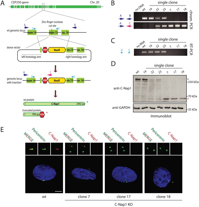
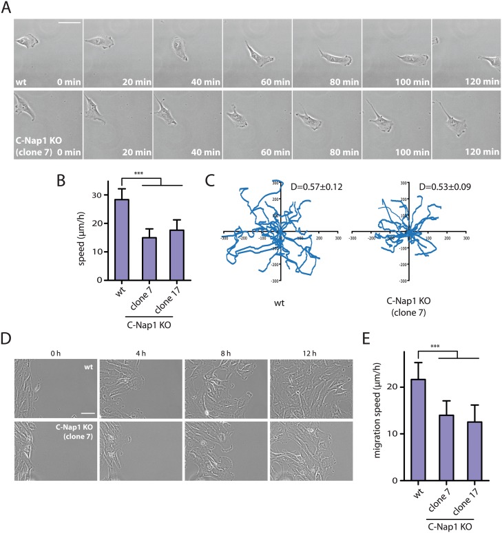
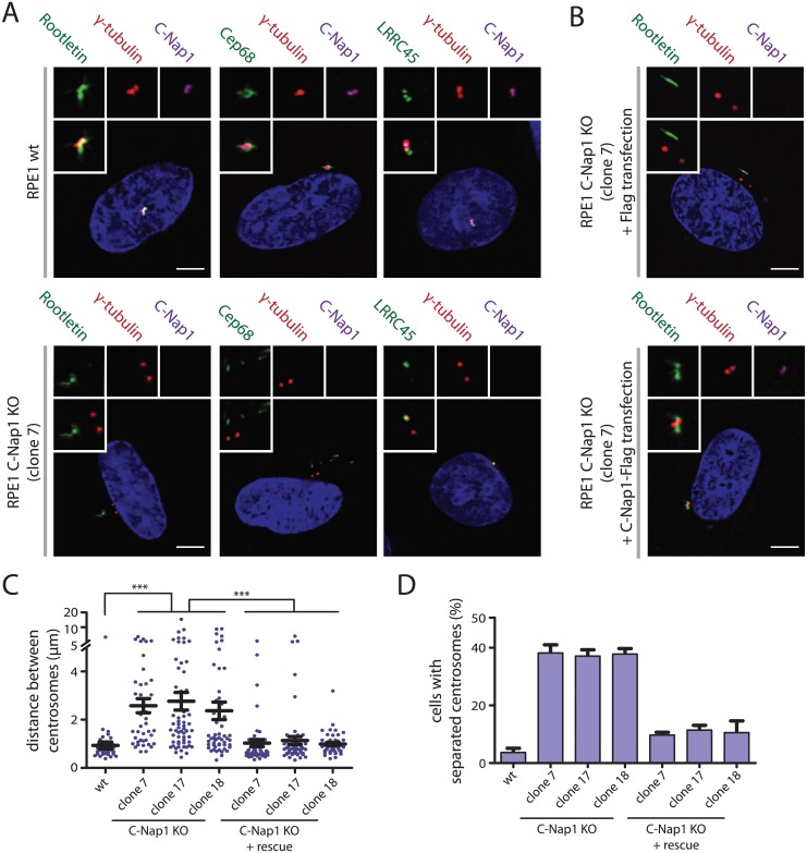
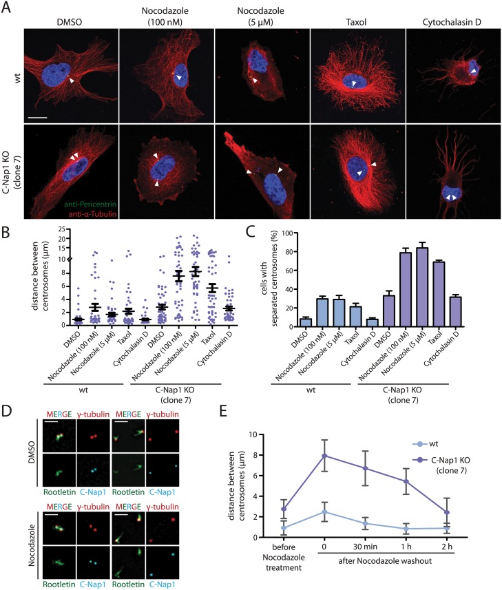
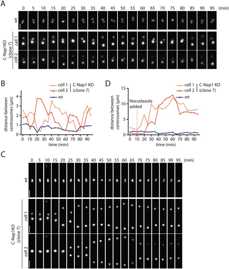
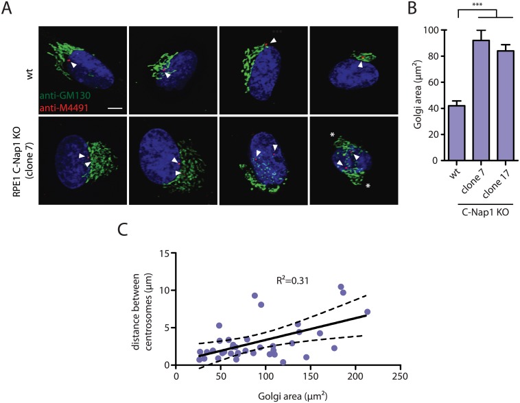
The centrosome is the principal microtubule organizing center in most animal cells. It consists of a pair of centrioles surrounded by pericentriolar material. The centrosome, like DNA, duplicates exactly once per cell cycle. During interphase duplicated centrosomes remain closely linked by a proteinaceous linker. This centrosomal linker is composed of rootletin filaments that are anchored to the centrioles via the protein C-Nap1. At the onset of mitosis the linker is dissolved by Nek2A kinase to support the formation of the bipolar mitotic spindle. The importance of the centrosomal linker for cell function during interphase awaits characterization. Here we assessed the phenotype of human RPE1 C-Nap1 knockout (KO) cells. The absence of the linker led to a modest increase in the average centrosome separation from 1 to 2.5 μm. This small impact on the degree of separation is indicative of a second level of spatial organization of centrosomes. Microtubule depolymerisation or stabilization in C-Nap1 KO cells dramatically increased the inter-centrosomal separation (> 8 μm). Thus, microtubules position centrosomes relatively close to one another in the absence of linker function. C-Nap1 KO cells had a Golgi organization defect with a two-fold expansion of the area occupied by the Golgi. When the centrosomes of C-Nap1 KO cells showed considerable separation, two spatially distinct Golgi stacks could be observed. Furthermore, migration of C-Nap1 KO cells was slower than their wild type RPE1 counterparts. These data show that the spatial organization of centrosomes is modulated by a combination of centrosomal cohesion and microtubule forces. Furthermore a modest increase in centrosome separation has major impact on Golgi organization and cell migration.
中心体是大多数动物细胞中主要的微管组织中心。它由一对中心粒和周围的中心粒周围物质组成。中心体如同DNA一样,在每个细胞周期精确复制一次。在间期,复制后的中心体通过一种蛋白质连接物保持紧密相连。这种中心体连接物由根蛋白细丝组成,这些细丝通过蛋白质C-Nap1锚定在中心粒上。在有丝分裂开始时,连接物被Nek2A激酶溶解,以支持双极有丝分裂纺锤体的形成。中心体连接物在间期对细胞功能的重要性尚待明确。在此,我们评估了人视网膜色素上皮(RPE1)细胞C-Nap1基因敲除(KO)后的表型。连接物的缺失导致中心体平均间距从1μm适度增加到2.5μm。这种对间距程度的微小影响表明中心体存在二级空间组织。在C-Nap1基因敲除细胞中,微管解聚或稳定会显著增加中心体间的间距(>8μm)。因此,在缺乏连接物功能时,微管将中心体相对紧密地定位在一起。C-Nap1基因敲除细胞存在高尔基体组织缺陷,高尔基体所占面积扩大了两倍。当C-Nap1基因敲除细胞的中心体表现出明显分离时,可以观察到两个空间上不同的高尔基体堆叠。此外,C-Nap1基因敲除细胞的迁移速度比野生型RPE1细胞慢。这些数据表明,中心体的空间组织受到中心体凝聚力和微管力的共同调节。此外,中心体间距的适度增加对高尔基体组织和细胞迁移有重大影响。