Jährling Nina, Becker Klaus, Wegenast-Braun Bettina M, Grathwohl Stefan A, Jucker Mathias, Dodt Hans-Ulrich
Department of Bioelectronics, FKE, Vienna University of Technology, Vienna, Austria; Section of Bioelectronics, Center for Brain Research (MUW), Vienna, Austria.
Department of Cellular Neurology, Hertie Institute for Clinical Brain Research, University of Tübingen, Tübingen, Germany; German Center for Neurodegenerative Diseases (DZNE), Tübingen, Germany.
PLoS One. 2015 May 27;10(5):e0125418. doi: 10.1371/journal.pone.0125418. eCollection 2015.
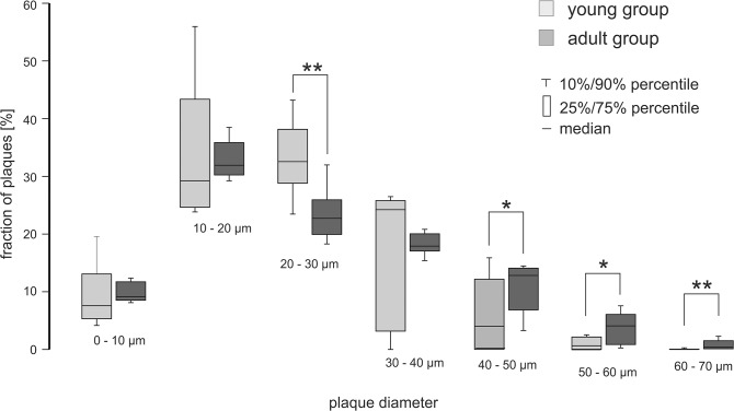
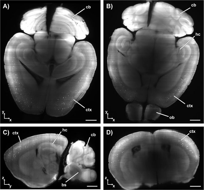
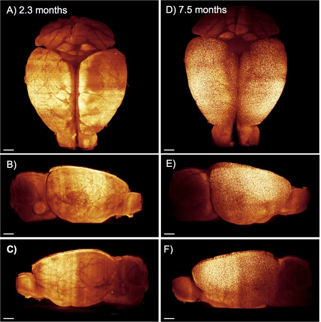
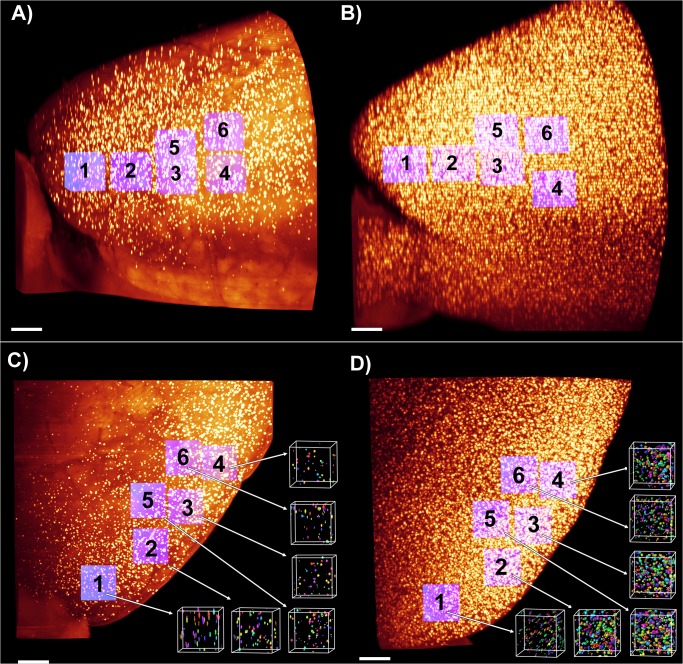
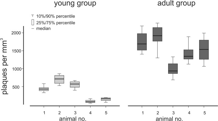
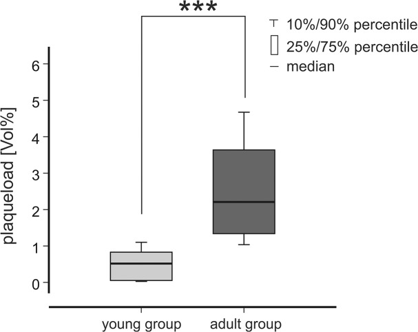
Alzheimer´s disease (AD) is the most common neurodegenerative disorder. AD neuropathology is characterized by intracellular neurofibrillary tangles and extracellular β-amyloid deposits in the brain. To elucidate the complexity of AD pathogenesis a variety of transgenic mouse models have been generated. An ideal imaging system for monitoring β-amyloid plaque deposition in the brain of these animals should allow 3D-reconstructions of β-amyloid plaques via a single scan of an uncropped brain. Ultramicroscopy makes this possible by replacing mechanical slicing in standard histology by optical sectioning. It allows a time efficient analysis of the amyloid plaque distribution in the entire mouse brain with 3D cellular resolution. We herein labeled β-amyloid deposits in a transgenic mouse model of cerebral β-amyloidosis (APPPS1 transgenic mice) with two intraperitoneal injections of the amyloid-binding fluorescent dye methoxy-X04. Upon postmortem analysis the total number of β-amyloid plaques, the β-amyloid load (volume percent) and the amyloid plaque size distributions were measured in the frontal cortex of two age groups (2.5 versus 7-8.5 month old mice). Applying ultramicroscopy we found in a proof-of-principle study that the number of β-amyloid plaques increases with age. In our experiments we further observed an increase of large plaques in the older age group of mice. We demonstrate that ultramicroscopy is a fast, and accurate analysis technique for studying β-amyloid lesions in transgenic mice allowing the 3D staging of β-amyloid plaque development. This in turn is the basis to study neural network degeneration upon cerebral β-amyloidosis and to assess Aβ-targeting therapeutics.
阿尔茨海默病(AD)是最常见的神经退行性疾病。AD神经病理学的特征是大脑中存在细胞内神经原纤维缠结和细胞外β-淀粉样蛋白沉积。为了阐明AD发病机制的复杂性,已经构建了多种转基因小鼠模型。一种理想的用于监测这些动物大脑中β-淀粉样蛋白斑块沉积的成像系统应能通过对未裁剪大脑的单次扫描实现β-淀粉样蛋白斑块的三维重建。超微显微镜通过用光学切片取代标准组织学中的机械切片实现了这一点。它能够以三维细胞分辨率对整个小鼠大脑中的淀粉样蛋白斑块分布进行高效分析。我们在此通过腹腔内注射两次淀粉样蛋白结合荧光染料甲氧基-X04,标记了脑β-淀粉样变性转基因小鼠模型(APPPS1转基因小鼠)中的β-淀粉样蛋白沉积物。在死后分析中,测量了两个年龄组(2.5个月与7 - 8.5个月大的小鼠)额叶皮质中β-淀粉样蛋白斑块的总数、β-淀粉样蛋白负荷(体积百分比)和淀粉样蛋白斑块大小分布。在一项原理验证研究中,应用超微显微镜我们发现β-淀粉样蛋白斑块的数量随年龄增加。在我们的实验中,我们还进一步观察到老年组小鼠中大斑块数量增加。我们证明超微显微镜是一种快速、准确的分析技术,可用于研究转基因小鼠中的β-淀粉样病变,实现β-淀粉样蛋白斑块发育的三维分期。这反过来又是研究脑β-淀粉样变性后神经网络退化以及评估靶向Aβ治疗方法的基础。