Lichtenegger Antonia, Muck Martina, Eugui Pablo, Harper Danielle J, Augustin Marco, Leskovar Konrad, Hitzenberger Christoph K, Woehrer Adelheid, Baumann Bernhard
Medical University of Vienna, Center for Medical Physics and Biomedical Engineering, Vienna, Austria.
General Hospital and Medical University of Vienna, Institute of Neurology, Vienna, Austria.
Neurophotonics. 2018 Jul;5(3):035002. doi: 10.1117/1.NPh.5.3.035002. Epub 2018 Jul 24.
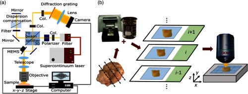
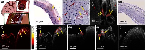
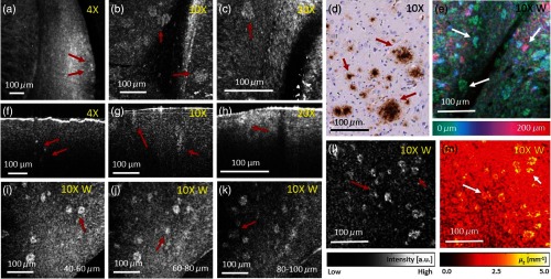
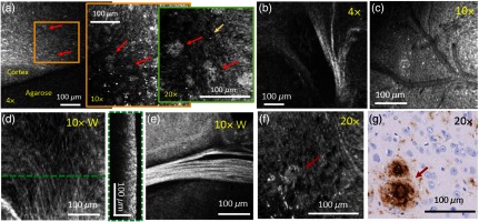
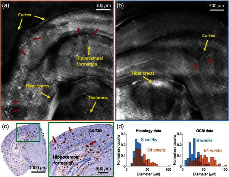
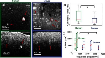
We implemented a wide field-of-view visible-light optical coherence microscope (OCM) for investigating brain tissue of patients diagnosed with Alzheimer's disease (AD) and of a mouse model of AD. A submicrometer axial resolution in tissue was achieved using a broad visible light spectrum. The use of various objective lenses enabled reaching micrometer transversal resolution and the acquisition of images of microscopic brain features, such as cell structures, vessels, and white matter tracts. Amyloid-beta plaques in the range of 10 to were visualized. Large field-of-view images of young and old mouse brain sections were imaged using an automated stage. The plaque load was characterized, revealing an age-related increase. Human brain tissue affected by cerebral amyloid angiopathy was investigated and hyperscattering structures resembling amyloid beta accumulations in the vessel walls were identified. All results were in good agreement with histology. A comparison of plaque features in both human and mouse brain tissue was performed, revealing an increase in plaque load and a decrease in reflectivity for mouse as compared with human brain tissue. Based on the promising outcome of our experiments, visible light OCM might be a powerful tool for investigating microscopic features in brain tissue.
我们开发了一种宽视野可见光光学相干显微镜(OCM),用于研究被诊断患有阿尔茨海默病(AD)的患者以及AD小鼠模型的脑组织。利用宽可见光谱在组织中实现了亚微米级轴向分辨率。使用各种物镜能够达到微米级横向分辨率,并获取微观脑特征的图像,如细胞结构、血管和白质束。可视化了10至 范围内的β淀粉样蛋白斑块。使用自动载物台对幼年和老年小鼠脑切片的大视野图像进行成像。对斑块负荷进行了表征,发现其与年龄相关增加。对受脑淀粉样血管病影响的人类脑组织进行了研究,并在血管壁中识别出类似于β淀粉样蛋白聚集的高散射结构。所有结果与组织学结果高度一致。对人类和小鼠脑组织中的斑块特征进行了比较,结果显示与人类脑组织相比,小鼠的斑块负荷增加且反射率降低。基于我们实验的良好结果,可见光OCM可能是研究脑组织微观特征的有力工具。