Department of Veterinary Sciences, University of Messina, Polo Universitario Annunziata, Messina, 98168, Italy.
Department of Veterinary Sciences, School of Agrarian and Veterinary Sciences, University of Trás-os-Montes e Alto Douro (UTAD), Vila Real, 5000-801, Portugal.
Parasit Vectors. 2015 Jun 4;8:302. doi: 10.1186/s13071-015-0909-z.
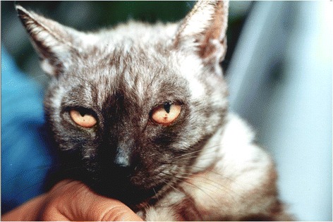
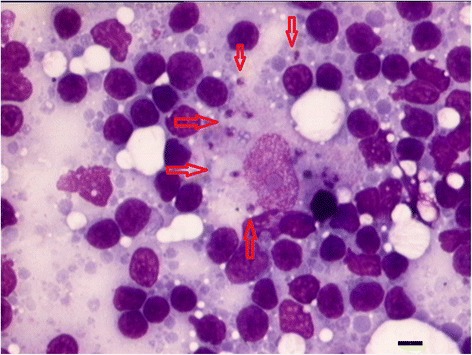
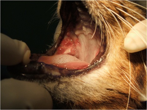
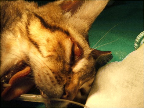
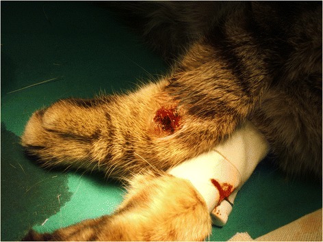
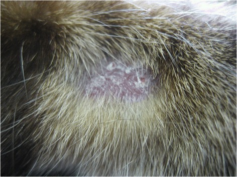
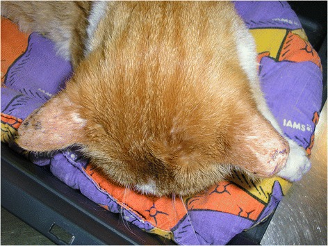
Limited data is available on feline leishmaniosis (FeL) caused by Leishmania infantum worldwide. The LeishVet group presents in this report a review of the current knowledge on FeL, the epidemiological role of the cat in L. infantum infection, clinical manifestations, and recommendations on diagnosis, treatment and monitoring, prognosis and prevention of infection, in order to standardize the management of this disease in cats. The consensus of opinions and recommendations was formulated by combining a comprehensive review of evidence-based studies and case reports, clinical experience and critical consensus discussions. While subclinical feline infections are common in areas endemic for canine leishmaniosis, clinical illness due to L. infantum in cats is rare. The prevalence rates of feline infection with L. infantum in serological or molecular-based surveys range from 0% to more than 60%. Cats are able to infect sand flies and, therefore, they may act as a secondary reservoir, with dogs being the primary natural reservoir. The most common clinical signs and clinicopathological abnormalities compatible with FeL include lymph node enlargement and skin lesions such as ulcerative, exfoliative, crusting or nodular dermatitis (mainly on the head or distal limbs), ocular lesions (mainly uveitis), feline chronic gingivostomatitis syndrome, mucocutaneous ulcerative or nodular lesions, hypergammaglobulinaemia and mild normocytic normochromic anaemia. Clinical illness is frequently associated with impaired immunocompetence, as in case of retroviral coinfections or immunosuppressive therapy. Diagnosis is based on serology, polymerase chain reaction (PCR), cytology, histology, immunohistochemistry (IHC) or culture. If serological testing is negative or low positive in a cat with clinical signs compatible with FeL, the diagnosis of leishmaniosis should not be excluded and additional diagnostic methods (cytology, histology with IHC, PCR, culture) should be employed. The most common treatment used is allopurinol. Meglumine antimoniate has been administered in very few reported cases. Both drugs are administered alone and most cats recover clinically after therapy. Follow-up of treated cats with routine laboratory tests, serology and PCR is essential for prevention of clinical relapses. Specific preventative measures for this infection in cats are currently not available.
关于全球范围内由利什曼原虫引起的猫利什曼病(FeL),可用的数据有限。LeishVet 小组在本报告中回顾了目前关于 FeL 的知识,包括猫在利什曼原虫感染中的流行病学作用、临床表现以及诊断、治疗和监测、预后和感染预防的建议,以便规范猫利什曼病的管理。意见和建议的共识是通过综合审查基于证据的研究和病例报告、临床经验和关键共识讨论来制定的。虽然亚临床猫感染在犬利什曼病流行地区很常见,但由于 L. infantum 引起的猫临床疾病很少见。在基于血清学或分子的调查中,猫感染 L. infantum 的流行率从 0%到超过 60%不等。猫能够感染沙蝇,因此它们可能是二次储主,而狗是主要的天然储主。最常见的与 FeL 相符的临床症状和临床病理异常包括淋巴结肿大和皮肤病变,如溃疡性、剥脱性、结痂或结节性皮炎(主要在头部或四肢末端)、眼部病变(主要是葡萄膜炎)、猫慢性牙龈炎综合征、黏膜溃疡或结节性病变、高丙种球蛋白血症和轻度正细胞正色素性贫血。临床疾病常与免疫功能受损有关,如逆转录病毒合并感染或免疫抑制治疗。诊断基于血清学、聚合酶链反应(PCR)、细胞学、组织学、免疫组织化学(IHC)或培养。如果具有 FeL 临床症状的猫的血清学检测结果为阴性或低阳性,则不应排除利什曼病的诊断,应采用其他诊断方法(细胞学、IHC 组织学、PCR、培养)。最常用的治疗药物是别嘌醇。在极少数报道的病例中使用了葡甲胺锑。这两种药物均单独使用,大多数猫在治疗后临床康复。对接受治疗的猫进行常规实验室检查、血清学和 PCR 随访对于预防临床复发至关重要。目前尚无针对猫这种感染的特定预防措施。