Diaz-Valdivia Natalia, Bravo Denisse, Huerta Hernán, Henriquez Soledad, Gabler Fernando, Vega Margarita, Romero Carmen, Calderon Claudia, Owen Gareth I, Leyton Lisette, Quest Andrew F G
Advanced Center for Chronic Diseases (ACCDiS), Santiago, Chile.
Center for Molecular studies of the Cell (CEMC), Programa de Biologia Celular y Molecular, Instituto de Ciencias Biomedicas, Facultad de Medicina, Universidad de Chile, Santiago, Chile.
BMC Cancer. 2015 Jun 10;15:463. doi: 10.1186/s12885-015-1477-5.
Caveolin-1 (CAV1) has been implicated both in tumor suppression and progression, whereby the specific role appears to be context dependent. Endometrial cancer is one of the most common malignancies of the female genital tract; however, little is known about the role of CAV1 in this disease.
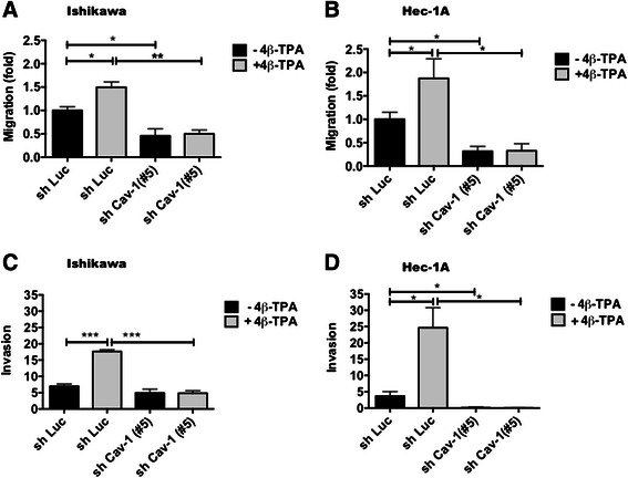
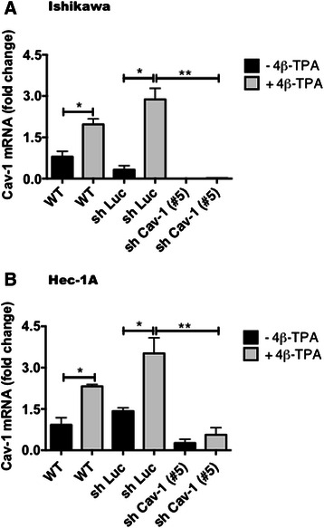
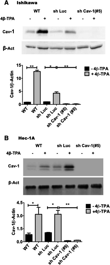
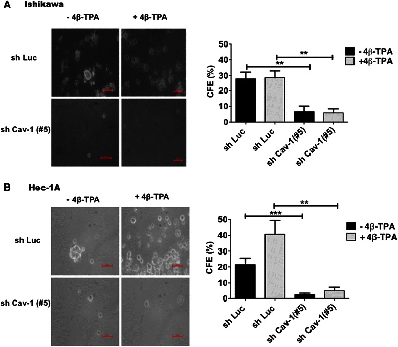
Here, we first determined by immunohistochemistry CAV1 protein levels in normal proliferative human endometrium and endometrial tumor samples. Then using two endometrial cancer cell lines (ECC: Ishikawa and Hec-1A) we evaluated mRNA and protein levels of CAV1 by real time qPCR and Western blot analysis, respectively. The role of CAV1 expression in ECC malignancy was further studied by either inducing its expression in endometrial cancer cells with the tumor promotor 12-O-tetradecanoyl-phorbol-13-acetate (4β-TPA) or decreasing expression using short-hairpin RNA constructs, and then evaluating the effects of these changes on ECC proliferation, transmigration, matrigel invasion, and colony formation in soft agar.
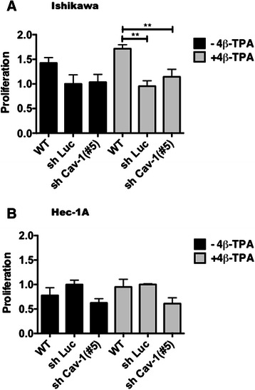
Immunohistochemical analysis of endometrial epithelia revealed that substantially higher levels of CAV1 were present in endometrial tumors than the normal proliferative epithelium. Also, in Ishikawa and Hec-1A endometrial cancer cells CAV1 expression was readily detectable. Upon treatment with 4β-TPA CAV1 levels increased and coincided with augmented cell transmigration, matrigel invasion, as well as colony formation in soft agar. Reduction of CAV1 expression using short-hairpin RNA constructs ablated these effects in both cell types whether treated or not with 4β-TPA. Alternatively, CAV1 expression appeared not to modulate significantly proliferation of these cells.
Our study shows that elevated CAV1, observed in patients with endometrial cancer, is linked to enhanced malignancy of endometrial cancer cells, as evidenced by increased migration, invasion and anchorage-independent growth.
小窝蛋白-1(CAV1)在肿瘤抑制和进展中均有涉及,但其具体作用似乎取决于环境。子宫内膜癌是女性生殖道最常见的恶性肿瘤之一;然而,关于CAV1在这种疾病中的作用知之甚少。
在此,我们首先通过免疫组织化学确定正常增殖期人子宫内膜和子宫内膜肿瘤样本中CAV1蛋白水平。然后使用两种子宫内膜癌细胞系(ECC:Ishikawa和Hec-1A),我们分别通过实时定量PCR和蛋白质印迹分析评估CAV1的mRNA和蛋白水平。通过用肿瘤促进剂12-O-十四烷酰佛波醇-13-乙酸酯(4β-TPA)诱导子宫内膜癌细胞中CAV1的表达或使用短发夹RNA构建体降低其表达,进一步研究CAV1表达在ECC恶性肿瘤中的作用,然后评估这些变化对ECC增殖、迁移、基质胶侵袭和软琼脂中集落形成的影响。
子宫内膜上皮的免疫组织化学分析显示,子宫内膜肿瘤中CAV1的水平明显高于正常增殖上皮。此外,在Ishikawa和Hec-1A子宫内膜癌细胞中很容易检测到CAV1的表达。用4β-TPA处理后,CAV1水平升高,并与细胞迁移、基质胶侵袭以及软琼脂中集落形成的增加同时出现。使用短发夹RNA构建体降低CAV1表达消除了这两种细胞类型中的这些效应,无论是否用4β-TPA处理。另外,CAV1表达似乎并未显著调节这些细胞的增殖。
我们的研究表明,在子宫内膜癌患者中观察到的CAV1升高与子宫内膜癌细胞恶性程度增加有关,这表现为迁移、侵袭和非锚定依赖性生长增加。