• 文献检索
  • 文档翻译
  • 深度研究
  • 学术资讯
  • Suppr Zotero 插件Zotero 插件
  • 邀请有礼
  • 套餐&价格
  • 历史记录
应用&插件
Suppr Zotero 插件Zotero 插件浏览器插件Mac 客户端Windows 客户端微信小程序
定价
高级版会员购买积分包购买API积分包
服务
文献检索文档翻译深度研究API 文档MCP 服务
关于我们
关于 Suppr公司介绍联系我们用户协议隐私条款
关注我们

Suppr 超能文献

核心技术专利:CN118964589B侵权必究
粤ICP备2023148730 号-1Suppr @ 2026

文献检索

告别复杂PubMed语法,用中文像聊天一样搜索,搜遍4000万医学文献。AI智能推荐,让科研检索更轻松。

立即免费搜索

文件翻译

保留排版,准确专业,支持PDF/Word/PPT等文件格式,支持 12+语言互译。

免费翻译文档

深度研究

AI帮你快速写综述,25分钟生成高质量综述,智能提取关键信息,辅助科研写作。

立即免费体验

内侧乳头肌复合体及其相关的隔缘肉柱。一项关于人类心脏的正常解剖学研究。

The medial papillary muscle complex and its related septomarginal trabeculation. A normal anatomical study on human hearts.

作者信息

Restivo A, Smith A, Wilkinson J L, Anderson R H

机构信息

Institute of Child Health, University of Liverpool, Royal Liverpool Children's Hospital Alder Hey.

出版信息

J Anat. 1989 Apr;163:231-42.

PMID:2606775
原文链接:https://pmc.ncbi.nlm.nih.gov/articles/PMC1256532/
Abstract

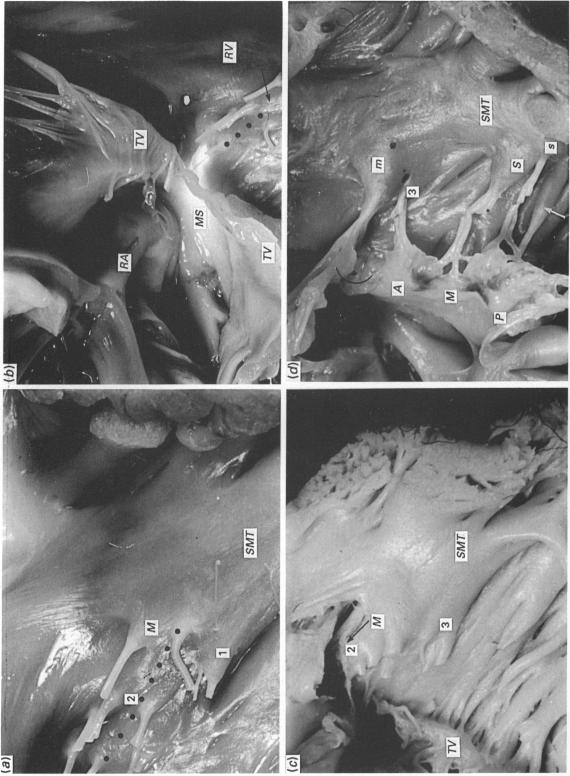
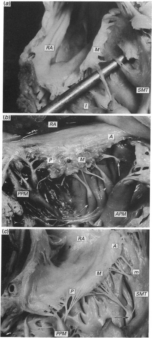
The morphology of the medial papillary muscle complex of the right ventricle was studied in 81 normal hearts from subjects ranging in age from 20 weeks of gestation to 13 months. The position of the main medial papillary muscle (of Lancisi) was differentiated in terms of a root and a belly. The anterior aspect of the root was found in a constant position at the basal bifurcation of the septomarginal trabeculation, whereas its belly showed considerable positional variation. Three groups of minor papillary muscles were identified and localised in specific areas in the base of the right ventricle. Taken overall, they formed part of the medial papillary complex. The complex itself was found to be constantly related to the antero-septal commissure of the tricuspid valve. A separate group of septal papillary muscles could be differentiated from the medial papillary complex. The anchorage of the septal leaflet of the tricuspid valve to the ventricular septum was also studied. The antero-superior and postero-inferior portions of this leaflet were found to be supported by the medial and posterior papillary complexes, respectively. Its midportion was connected to the septal group of papillary muscles but much variability was evident. The portion of the septomarginal trabeculation supporting the medial papillary complex, namely the postero-basal division, was studied further. This showed considerable variability and did not form a continuous anatomical spectrum.

摘要

对81例年龄从妊娠20周至13个月的正常心脏的右心室内侧乳头肌复合体形态进行了研究。主要内侧乳头肌(兰西乳头肌)的位置根据根部和肌腹进行区分。根部的前侧位于隔缘小梁基部的恒定位置,而其肌腹位置变化较大。确定了三组小乳头肌并将其定位在右心室基部的特定区域。总体而言,它们构成了内侧乳头肌复合体的一部分。发现该复合体与三尖瓣的前间隔连合始终相关。可以从内侧乳头肌复合体中区分出一组单独的间隔乳头肌。还研究了三尖瓣隔叶与室间隔的附着情况。发现该叶的前上部分和后下部分分别由内侧和后侧乳头肌复合体支撑。其中部与间隔乳头肌群相连,但变异明显。对支撑内侧乳头肌复合体的隔缘小梁部分,即后基部进行了进一步研究。结果显示其变异较大,未形成连续的解剖谱系。

https://cdn.ncbi.nlm.nih.gov/pmc/blobs/c2fa/1256532/0d620a88b90f/janat00047-0235-a.jpg
https://cdn.ncbi.nlm.nih.gov/pmc/blobs/c2fa/1256532/b9aa6ba8369c/janat00047-0229-a.jpg
https://cdn.ncbi.nlm.nih.gov/pmc/blobs/c2fa/1256532/bf2071c8efcb/janat00047-0231-a.jpg
https://cdn.ncbi.nlm.nih.gov/pmc/blobs/c2fa/1256532/e647b56b6ed5/janat00047-0234-a.jpg
https://cdn.ncbi.nlm.nih.gov/pmc/blobs/c2fa/1256532/0d620a88b90f/janat00047-0235-a.jpg

相似文献

1
The medial papillary muscle complex and its related septomarginal trabeculation. A normal anatomical study on human hearts.内侧乳头肌复合体及其相关的隔缘肉柱。一项关于人类心脏的正常解剖学研究。
J Anat. 1989 Apr;163:231-42.
2
Normal variations in the relationship of the tricuspid valve to the membranous septum in the human heart.人心脏中三尖瓣与膜性间隔关系的正常变异。
Anat Rec. 1990 Feb;226(2):258-63. doi: 10.1002/ar.1092260215.
3
Anatomy of the septomarginal trabecula in Landrace pig hearts.
Morphologie. 2010 May;94(305):26-9. doi: 10.1016/j.morpho.2010.03.004. Epub 2010 Mar 31.
4
Anatomy of the septomarginal trabecula in goat hearts.
Ital J Anat Embryol. 2010;115(3):229-34.
5
Remarks on the morphology of the papillary muscles of the right ventricle.关于右心室乳头肌形态学的论述
Folia Morphol (Warsz). 2005 Aug;64(3):176-82.
6
Positions of septal papillary muscles in human hearts.
Folia Morphol (Warsz). 2010 May;69(2):101-6.
7
Variations in the papillary muscles of normal tricuspid valve and their clinical relevance in medicolegal autopsies.正常三尖瓣乳头肌的变异及其在法医尸检中的临床意义。
Saudi Med J. 2004 Sep;25(9):1176-85.
8
Provision of tricuspid valve leaflets by septal papillary muscles in the right ventricle of human and other mammal hearts.
Folia Morphol (Warsz). 2003;62(3):309-11.
9
Assessment of the anatomical variation to be found in the normal tricuspid valve.对正常三尖瓣解剖变异的评估。
Clin Anat. 2016 Apr;29(3):399-407. doi: 10.1002/ca.22591. Epub 2015 Jul 16.
10
An endoscopic and anatomical approach to the septal papillary muscle of the conus.一种针对圆锥间隔乳头肌的内镜及解剖学入路。
Surg Radiol Anat. 2009 Nov;31(9):701-6. doi: 10.1007/s00276-009-0510-2. Epub 2009 May 5.

引用本文的文献

1
Usefulness of the "En face view" method for diagnosing perimembranous ventricular septal defects.“正面观”法在诊断膜周部室间隔缺损中的应用价值。
J Echocardiogr. 2024 Dec 28. doi: 10.1007/s12574-024-00678-4.
2
Congenitally corrected transposition of the great arteries: is it really a transposition? An anatomical study of the right ventricular septal surface.先天性矫正性大动脉转位:它真的是转位吗?对右心室间隔面的解剖学研究。
J Anat. 2020 Feb;236(2):325-333. doi: 10.1111/joa.13097. Epub 2019 Oct 27.
3
Tricuspid and Pulmonic Valve Pathology.

本文引用的文献

1
The medial papillary complex.内侧乳头复合体
Br Heart J. 1977 Sep;39(9):1012-8. doi: 10.1136/hrt.39.9.1012.
三尖瓣和肺动脉瓣病变。
Curr Cardiol Rep. 2019 May 18;21(7):54. doi: 10.1007/s11886-019-1143-7.
4
Right ventricular septomarginal trabeculation hypertrophy is associated with disease severity in patients with pulmonary arterial hypertension.右心室隔缘小梁肥厚与肺动脉高压患者的疾病严重程度相关。
Int J Cardiovasc Imaging. 2018 Sep;34(9):1439-1449. doi: 10.1007/s10554-018-1347-8. Epub 2018 Mar 31.
5
The trabecula septomarginalis (Leonardo's cord) in abnormal ventriculo-arterial connections: anatomic and morphogenetic implications.异常心室-动脉连接中的隔缘小梁(列奥纳多氏索):解剖学及形态发生学意义
J Cardiothorac Surg. 2014 Apr 21;9:71. doi: 10.1186/1749-8090-9-71.
6
Endocavitary structures in the outflow tract: anatomy and electrophysiology of the conus papillary muscles.心腔流出道中的心内膜下结构:圆锥乳头肌的解剖与电生理学。
J Cardiovasc Electrophysiol. 2014 Jan;25(1):94-8. doi: 10.1111/jce.12291. Epub 2013 Oct 10.
7
Morphogenetic aspects of the septomarginal trabecula in the human heart.人类心脏中隔缘小梁的形态发生学方面。
Arch Med Sci. 2010 Oct;6(5):733-43. doi: 10.5114/aoms.2010.17089. Epub 2010 Oct 26.
8
Right ventricular variants and pulmonary embolism-association or coincidence?右心室变异与肺栓塞——关联还是巧合?
Clin Med Insights Cardiol. 2010 Jul 21;4:53-7. doi: 10.4137/cmc.s5244.
9
An endoscopic and anatomical approach to the septal papillary muscle of the conus.一种针对圆锥间隔乳头肌的内镜及解剖学入路。
Surg Radiol Anat. 2009 Nov;31(9):701-6. doi: 10.1007/s00276-009-0510-2. Epub 2009 May 5.
10
Embryogenesis of the heart muscle.心肌的胚胎发生。
Heart Fail Clin. 2008 Jul;4(3):235-45. doi: 10.1016/j.hfc.2008.02.007.