• 文献检索
  • 文档翻译
  • 深度研究
  • 学术资讯
  • Suppr Zotero 插件Zotero 插件
  • 邀请有礼
  • 套餐&价格
  • 历史记录
应用&插件
Suppr Zotero 插件Zotero 插件浏览器插件Mac 客户端Windows 客户端微信小程序
定价
高级版会员购买积分包购买API积分包
服务
文献检索文档翻译深度研究API 文档MCP 服务
关于我们
关于 Suppr公司介绍联系我们用户协议隐私条款
关注我们

Suppr 超能文献

核心技术专利:CN118964589B侵权必究
粤ICP备2023148730 号-1Suppr @ 2026

文献检索

告别复杂PubMed语法,用中文像聊天一样搜索,搜遍4000万医学文献。AI智能推荐,让科研检索更轻松。

立即免费搜索

文件翻译

保留排版,准确专业,支持PDF/Word/PPT等文件格式,支持 12+语言互译。

免费翻译文档

深度研究

AI帮你快速写综述,25分钟生成高质量综述,智能提取关键信息,辅助科研写作。

立即免费体验

关节软骨中 T1ρ 弥散:与材料特性和大分子含量的关系。

T1ρ Dispersion in Articular Cartilage: Relationship to Material Properties and Macromolecular Content.

机构信息

Department of Mechanical Engineering, Stanford University, Stanford, CA, USA.

Auckland Bioengineering Institute, University of Auckland, Auckland, New Zealand.

出版信息

Cartilage. 2015 Apr;6(2):113-22. doi: 10.1177/1947603515569529.

DOI:10.1177/1947603515569529
PMID:26069714
原文链接:https://pmc.ncbi.nlm.nih.gov/articles/PMC4462251/
Abstract

OBJECTIVE

This study assessed T1ρ relaxation dispersion, measured by magnetic resonance imaging (MRI), as a tool to noninvasively evaluate cartilage material and biochemical properties. The specific objective was to answer two questions: (1) does cartilage initial elastic modulus (E 0) correlate with T1ρ dispersion effects and (2) does collagen or proteoglycan content correlate with T1ρ dispersion effects?

DESIGN

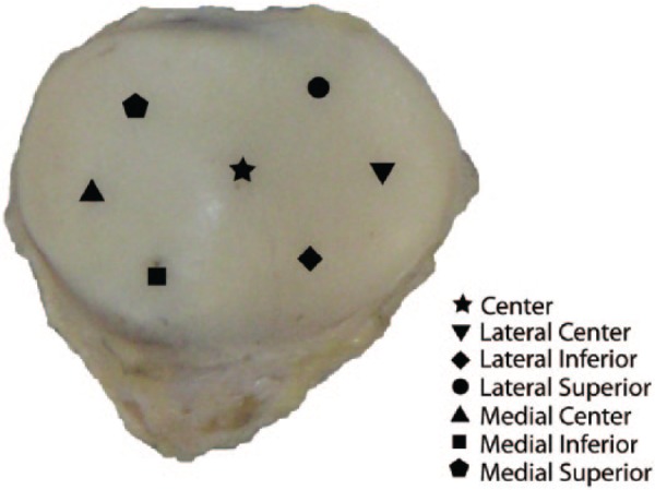
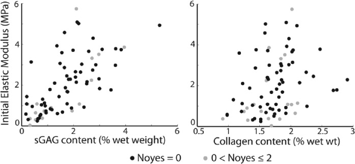
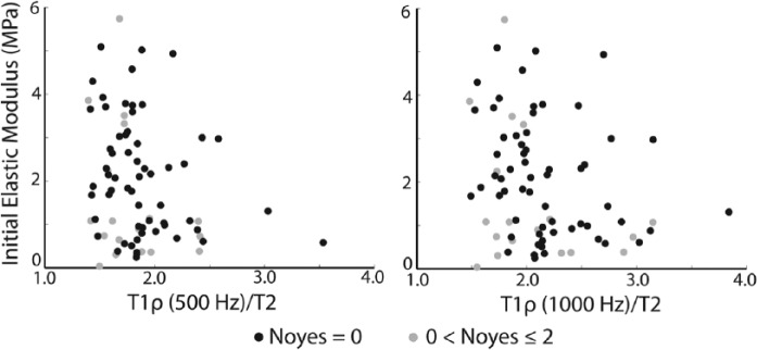
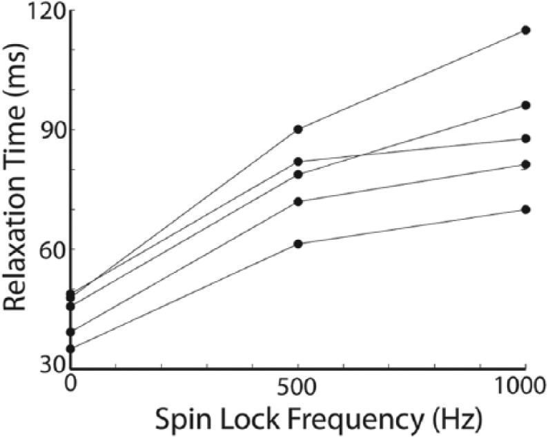
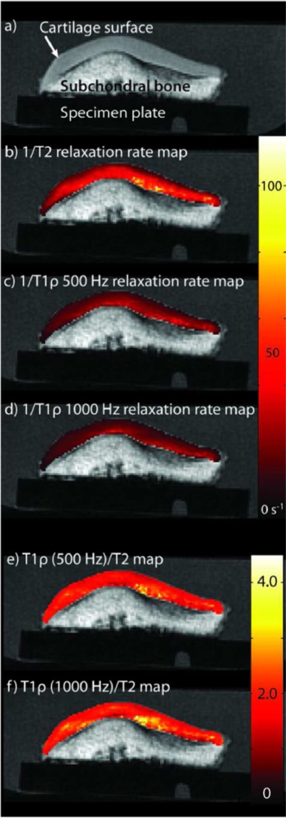
Cadaveric patellae with and without visible cartilage damage on conventional MR were included. T2 and T1ρ relaxation times at 500 and 1000 Hz spin-lock field amplitudes were measured. We estimated T1ρ dispersion effects by measuring T1ρ relaxation time at 500 and 1000 Hz and T2 relaxation time and using a new tool, the ratio T1ρ/T2. Cartilage initial elastic modulus, E 0, was measured from initial response of mechanical indentation creep tests. Collagen and proteoglycan contents were measured at the indentation test sites; proteoglycan content was measured by their covalently linked sulfated glycosaminoglycans (sGAG). Pearson correlation coefficients were determined, taking into account the clustering of multiple samples within a single patella specimen.

RESULTS

Cartilage initial elastic modulus, E 0, increased with decreasing values of T1ρ/T2 measurements at both 500 Hz (P = 0.034) and 1000 Hz (P = 0.022). 1/T1ρ relaxation time (500 Hz) increased with increasing sGAG content (P = 0.041).

CONCLUSIONS

T1ρ/T2 ratio, a new tool, and cartilage initial elastic modulus are both measures of water-protein interactions, are dependent on the cartilage structure, and were correlated in this study.

摘要

目的

本研究通过磁共振成像(MRI)评估 T1ρ弛豫弥散,以无创评估软骨的物质和生化特性。具体目的是回答两个问题:(1)软骨初始弹性模量(E0)是否与 T1ρ弥散效应相关;(2)胶原或蛋白聚糖含量是否与 T1ρ弥散效应相关。

设计

纳入常规 MRI 有或无可见软骨损伤的髌骨关节。测量 500Hz 和 1000Hz 自旋锁定场幅度下的 T2 和 T1ρ弛豫时间。我们通过测量 500Hz 和 1000Hz 的 T1ρ弛豫时间、T2 弛豫时间,并使用新工具 T1ρ/T2 比来估计 T1ρ 弥散效应。软骨初始弹性模量 E0 是通过机械压痕蠕变试验的初始响应来测量的。在压痕试验部位测量胶原和蛋白聚糖含量;蛋白聚糖含量通过其共价连接的硫酸化糖胺聚糖(sGAG)进行测量。考虑到单个髌骨关节标本内多个样本的聚类,确定了 Pearson 相关系数。

结果

在 500Hz(P = 0.034)和 1000Hz(P = 0.022)时,T1ρ/T2 测量值越低,软骨初始弹性模量 E0 越高。1/T1ρ 弛豫时间(500Hz)与 sGAG 含量呈正相关(P = 0.041)。

结论

T1ρ/T2 比,一种新的工具,以及软骨初始弹性模量都是水-蛋白相互作用的测量指标,依赖于软骨结构,在本研究中是相关的。

https://cdn.ncbi.nlm.nih.gov/pmc/blobs/4aff/4462251/001d721bbd4b/10.1177_1947603515569529-fig5.jpg
https://cdn.ncbi.nlm.nih.gov/pmc/blobs/4aff/4462251/2a35b6a78332/10.1177_1947603515569529-fig1.jpg
https://cdn.ncbi.nlm.nih.gov/pmc/blobs/4aff/4462251/bcdf235c5a84/10.1177_1947603515569529-fig2.jpg
https://cdn.ncbi.nlm.nih.gov/pmc/blobs/4aff/4462251/f4f06da7cec7/10.1177_1947603515569529-fig3.jpg
https://cdn.ncbi.nlm.nih.gov/pmc/blobs/4aff/4462251/6afed77e141b/10.1177_1947603515569529-fig4.jpg
https://cdn.ncbi.nlm.nih.gov/pmc/blobs/4aff/4462251/001d721bbd4b/10.1177_1947603515569529-fig5.jpg

相似文献

1
T1ρ Dispersion in Articular Cartilage: Relationship to Material Properties and Macromolecular Content.关节软骨中 T1ρ 弥散:与材料特性和大分子含量的关系。
Cartilage. 2015 Apr;6(2):113-22. doi: 10.1177/1947603515569529.
2
Relationship between T1rho magnetic resonance imaging, synovial fluid biomarkers, and the biochemical and biomechanical properties of cartilage.T1rho磁共振成像、滑液生物标志物与软骨生化及生物力学特性之间的关系。
J Biomech. 2017 Apr 11;55:18-26. doi: 10.1016/j.jbiomech.2017.02.001. Epub 2017 Feb 6.
3
Regional variation in T1ρ and T2 times in osteoarthritic human menisci: correlation with mechanical properties and matrix composition.骨关节炎人半月板 T1ρ 和 T2 值的区域性变化:与机械性能和基质组成的相关性。
Osteoarthritis Cartilage. 2013 Jun;21(6):796-805. doi: 10.1016/j.joca.2013.03.002. Epub 2013 Mar 14.
4
Prediction of glycosaminoglycan content in human cartilage by age, T1ρ and T2 MRI.基于年龄、T1ρ 和 T2 MRI 预测人软骨中的糖胺聚糖含量。
Osteoarthritis Cartilage. 2011 Feb;19(2):171-9. doi: 10.1016/j.joca.2010.11.009. Epub 2010 Nov 26.
5
Proteoglycan depletion-induced changes in transverse relaxation maps of cartilage: comparison of T2 and T1rho.蛋白聚糖耗竭引起的软骨横向弛豫图变化:T2与T1rho的比较
Acad Radiol. 2002 Dec;9(12):1388-94. doi: 10.1016/s1076-6332(03)80666-9.
6
T1rho-relaxation in articular cartilage: effects of enzymatic degradation.关节软骨中的T1rho弛豫:酶促降解的影响
Magn Reson Med. 1997 Dec;38(6):863-7. doi: 10.1002/mrm.1910380602.
7
Orientation anisotropy of quantitative MRI parameters in degenerated human articular cartilage.退变人关节软骨定量MRI参数的取向各向异性
J Orthop Res. 2021 Apr;39(4):861-870. doi: 10.1002/jor.24778. Epub 2020 Jun 29.
8
Bi-component T2 mapping correlates with articular cartilage material properties.双组分T2映射与关节软骨材料特性相关。
J Biomech. 2021 Feb 12;116:110215. doi: 10.1016/j.jbiomech.2020.110215. Epub 2020 Dec 31.
9
Assessing post-traumatic changes in cartilage using T dispersion parameters.使用T扩散参数评估软骨创伤后变化。
Magn Reson Imaging. 2023 Apr;97:91-101. doi: 10.1016/j.mri.2022.12.012. Epub 2023 Jan 4.
10
Topographical Variation of Human Femoral Articular Cartilage Thickness, T1rho and T2 Relaxation Times Is Related to Local Loading during Walking.人股骨关节软骨厚度、T1rho 和 T2 弛豫时间的地形变化与行走时的局部负荷有关。
Cartilage. 2019 Apr;10(2):229-237. doi: 10.1177/1947603517752057. Epub 2018 Jan 11.

引用本文的文献

1
SPICY: a method for single scan rotating frame relaxometry.SPICY:一种单扫描旋转框架弛豫测量法。
Phys Chem Chem Phys. 2023 May 10;25(18):13164-13169. doi: 10.1039/d2cp05988f.
2
New methods for robust continuous wave T relaxation preparation.用于稳健连续波 T1 弛豫制备的新方法。
NMR Biomed. 2023 Feb;36(2):e4834. doi: 10.1002/nbm.4834. Epub 2022 Oct 7.
3
Embedded Quantitative MRI T Mapping Using Non-Linear Primal-Dual Proximal Splitting.使用非线性原始对偶近端分裂的嵌入式定量磁共振成像T映射

本文引用的文献

1
A viscoelastic constitutive model can accurately represent entire creep indentation tests of human patella cartilage.粘弹性本构模型可以准确地呈现人体髌骨软骨的整个蠕变压痕试验。
J Appl Biomech. 2013 Jun;29(3):292-302. doi: 10.1123/jab.29.3.292. Epub 2012 Oct 1.
2
Orientational dependent sensitivities of T2 and T1ρ towards trypsin degradation and Gd-DTPA2- presence in bovine nasal cartilage.牛鼻软骨中胰蛋白酶降解和 Gd-DTPA2-存在对 T2 和 T1ρ 各向异性敏感性的影响。
MAGMA. 2012 Aug;25(4):297-304. doi: 10.1007/s10334-011-0288-1. Epub 2011 Nov 10.
3
T1ρ and T2 mapping reveal the in vivo extracellular matrix of articular cartilage.
J Imaging. 2022 May 31;8(6):157. doi: 10.3390/jimaging8060157.
4
Bi-component T2 mapping correlates with articular cartilage material properties.双组分T2映射与关节软骨材料特性相关。
J Biomech. 2021 Feb 12;116:110215. doi: 10.1016/j.jbiomech.2020.110215. Epub 2020 Dec 31.
5
T-based fibril-reinforced poroviscoelastic constitutive relation of human articular cartilage using inverse finite element technology.基于T的人关节软骨原纤维增强多孔粘弹性本构关系的逆有限元技术研究
Quant Imaging Med Surg. 2019 Mar;9(3):359-370. doi: 10.21037/qims.2019.03.01.
6
Relationship Between Quantitative MRI Biomarkers and Patient-Reported Outcome Measures After Cartilage Repair Surgery: A Systematic Review.软骨修复手术后定量MRI生物标志物与患者报告结局指标之间的关系:一项系统评价
Orthop J Sports Med. 2018 Apr 10;6(4):2325967118765448. doi: 10.1177/2325967118765448. eCollection 2018 Apr.
7
Experimental Influences in the Accurate Measurement of Cartilage Thickness in MRI.MRI 中软骨厚度精确测量的实验影响。
Cartilage. 2019 Jul;10(3):278-287. doi: 10.1177/1947603517749917. Epub 2018 Jan 3.
8
A non-contrast CMR index for assessing myocardial fibrosis.一种用于评估心肌纤维化的非对比心脏磁共振成像指标。
Magn Reson Imaging. 2017 Oct;42:69-73. doi: 10.1016/j.mri.2017.04.012. Epub 2017 Apr 28.
9
Composite metric R  - R (1/T  - 1/T ) as a potential MR imaging biomarker associated with changes in pain after ACL reconstruction: A six-month follow-up.复合指标R - R(1/T - 1/T)作为与前交叉韧带重建术后疼痛变化相关的潜在磁共振成像生物标志物:六个月随访。
J Orthop Res. 2017 Mar;35(3):718-729. doi: 10.1002/jor.23400. Epub 2016 Sep 16.
10
T1ρ magnetic resonance: basic physics principles and applications in knee and intervertebral disc imaging.T1ρ磁共振成像:基本物理原理及其在膝关节和椎间盘成像中的应用
Quant Imaging Med Surg. 2015 Dec;5(6):858-85. doi: 10.3978/j.issn.2223-4292.2015.12.06.
T1ρ 和 T2 映射显示关节软骨的细胞外基质的体内状态。
J Magn Reson Imaging. 2012 Jan;35(1):147-55. doi: 10.1002/jmri.22811. Epub 2011 Oct 11.
4
Local tissue properties of human osteoarthritic cartilage correlate with magnetic resonance T(1) rho relaxation times.人骨关节炎软骨的局部组织特性与磁共振 T1ρ弛豫时间相关。
J Orthop Res. 2011 Sep;29(9):1312-9. doi: 10.1002/jor.21381. Epub 2011 Mar 28.
5
Cross-relaxation imaging of human articular cartilage.人体关节软骨的交叉弛豫成像。
Magn Reson Med. 2011 Sep;66(3):725-34. doi: 10.1002/mrm.22865. Epub 2011 Mar 17.
6
Knee articular cartilage damage in osteoarthritis: analysis of MR image biomarker reproducibility in ACRIN-PA 4001 multicenter trial.骨关节炎膝关节关节软骨损伤:ACRIN-PA 4001 多中心试验中磁共振成像生物标志物重现性分析。
Radiology. 2011 Mar;258(3):832-42. doi: 10.1148/radiol.10101174. Epub 2011 Jan 6.
7
Quantitative MRI using T1ρ and T2 in human osteoarthritic cartilage specimens: correlation with biochemical measurements and histology.使用 T1ρ 和 T2 定量 MRI 检测人骨关节炎软骨标本:与生化测量和组织学的相关性。
Magn Reson Imaging. 2011 Apr;29(3):324-34. doi: 10.1016/j.mri.2010.09.004. Epub 2010 Dec 3.
8
Prediction of glycosaminoglycan content in human cartilage by age, T1ρ and T2 MRI.基于年龄、T1ρ 和 T2 MRI 预测人软骨中的糖胺聚糖含量。
Osteoarthritis Cartilage. 2011 Feb;19(2):171-9. doi: 10.1016/j.joca.2010.11.009. Epub 2010 Nov 26.
9
Combined off-resonance imaging and T2 relaxation in the rotating frame for positive contrast MR imaging of infection in a murine burn model.旋转框架下的离共振成像与 T2 弛豫联合用于鼠烧伤模型感染的正对比 MRI 成像。
J Magn Reson Imaging. 2010 Nov;32(5):1172-83. doi: 10.1002/jmri.22349.
10
T1rho MRI quantification of arthroscopically confirmed cartilage degeneration.关节镜确认的软骨退变的 T1rho MRI 定量分析。
Magn Reson Med. 2010 May;63(5):1376-82. doi: 10.1002/mrm.22272.