Brea David, Agulla Jesús, Staes An, Gevaert Kris, Campos Francisco, Sobrino Tomás, Blanco Miguel, Dávalos Antoni, Castillo José, Ramos-Cabrer Pedro
1] Neurology Department, Neurovascular Area, Clinical Neurosciences Research Laboratory, University Clinical Hospital, Health Research Institute of Santiago de Compostela (IDIS), University of Santiago de Compostela, Spain [2] Cellular and Molecular Neurobiology Research Group and Grup de Recerça en Neurociencies del IGTP, Department of Neurosciences, Fundació Institut d'Investigació en Ciències de la Salut Germans Trias I Pujol-Universitat Autónoma de Barcelona, Badalona, Spain.
1] Neurology Department, Neurovascular Area, Clinical Neurosciences Research Laboratory, University Clinical Hospital, Health Research Institute of Santiago de Compostela (IDIS), University of Santiago de Compostela, Spain [2] Research Unit, University Hospital of Salamanca and Institute of Health Sciences of Castilla and Leon, Salamanca, Spain.
Sci Rep. 2015 Jul 8;5:12030. doi: 10.1038/srep12030.
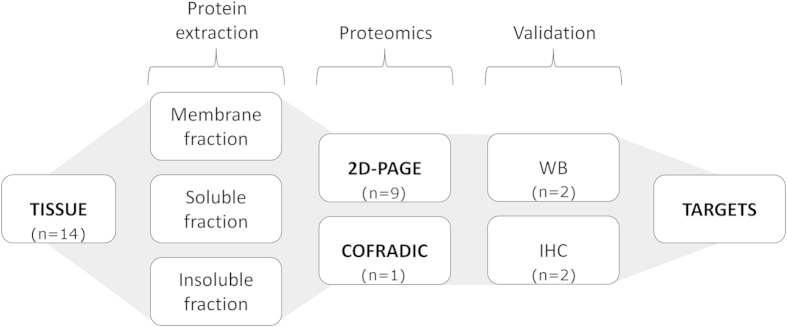
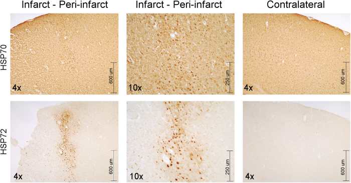
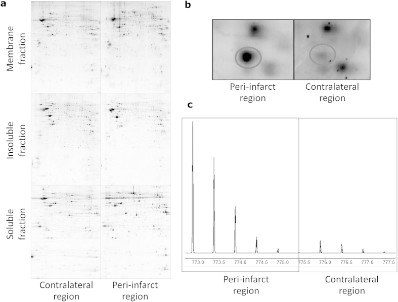
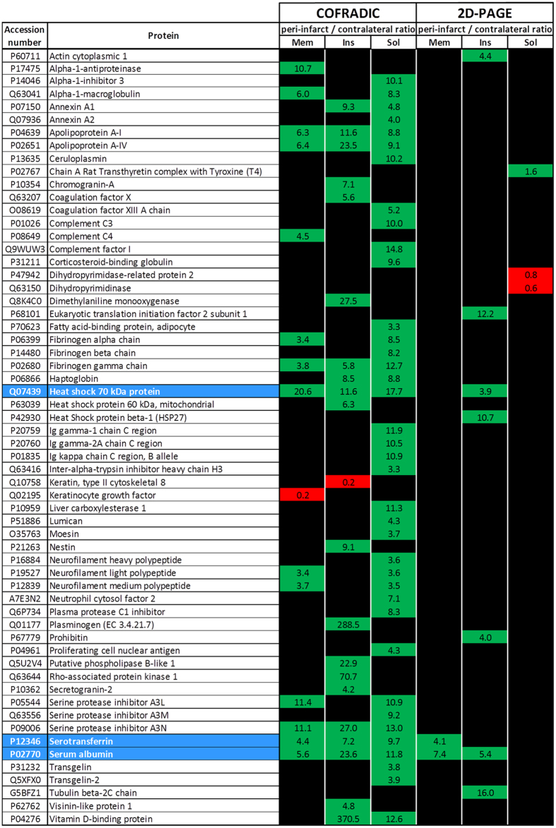
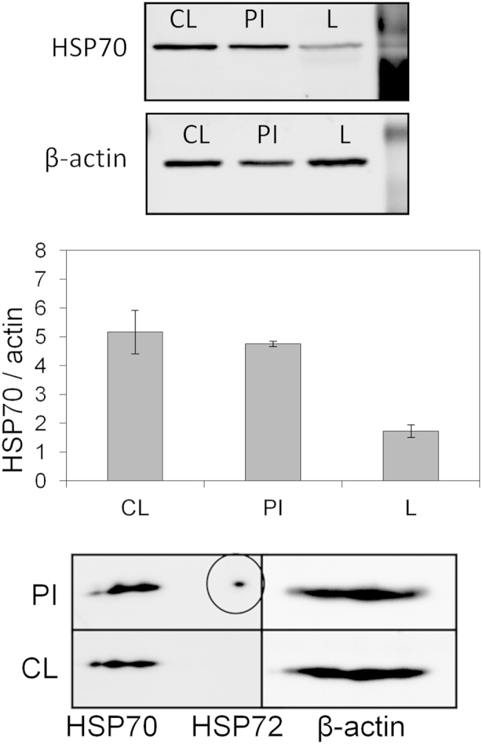
In this work, we report our study of protein expression in rat peri-infarct tissue, 48 h after the induction of permanent focal cerebral ischemia. Two proteomic approaches, gel electrophoresis with mass spectrometry and combined fractional diagonal chromatography (COFRADIC), were performed using tissue samples from the periphery of the induced cerebral ischemic lesions, using tissue from the contra-lateral hemisphere as a control. Several protein spots (3408) were identified by gel electrophoresis, and 11 showed significant differences in expression between peri-infarct and contra-lateral tissues (at least 3-fold, p < 0.05). Using COFRADIC, 5412 proteins were identified, with 72 showing a difference in expression. Apart from blood-related proteins (such as serum albumin), both techniques showed that the 70 kDa family of heat shock proteins were highly expressed in the peri-infarct tissue. Further studies by 1D and 2D western blotting and immunohistochemistry revealed that only one member of this family (the inducible form, HSP72 or HSP70i) is specifically expressed by the peri-infarct tissue, while the majority of this family (the constitutive form, HSC70 or HSP70c) is expressed in the whole brain. Our data support that HSP72 is a suitable biomarker of peri-infarct tissue in the ischemic brain.
在本研究中,我们报告了在永久性局灶性脑缺血诱导后48小时,对大鼠梗死周边组织中蛋白质表达的研究。我们采用了两种蛋白质组学方法,即凝胶电泳结合质谱分析以及组合分数对角线色谱法(COFRADIC),使用诱导性脑缺血损伤周边的组织样本,并以对侧半球的组织作为对照。通过凝胶电泳鉴定出了多个蛋白质斑点(3408个),其中11个在梗死周边组织和对侧组织之间表现出显著的表达差异(至少3倍,p<0.05)。使用COFRADIC方法,鉴定出了5412种蛋白质,其中72种表现出表达差异。除了与血液相关的蛋白质(如血清白蛋白)外,两种技术均显示70 kDa热休克蛋白家族在梗死周边组织中高表达。通过一维和二维蛋白质印迹法以及免疫组织化学的进一步研究表明,该家族中只有一个成员(诱导型,HSP72或HSP70i)在梗死周边组织中特异性表达,而该家族的大多数成员(组成型,HSC70或HSP70c)在全脑中表达。我们的数据支持HSP72是缺血性脑中梗死周边组织的合适生物标志物。