Meireles Diogo de Abreu, Schripsema Jan, Arnholdt Andrea Cristina Vetö, Dagnino Denise
Laboratório de Biotecnologia, Universidade Estadual do Norte Fluminense Darcy Ribeiro, Campos dos Goytacazes, Rio de Janeiro, Brazil.
Grupo Metabolômica, Laboratório de Ciências Químicas, Universidade Estadual do Norte Fluminense Darcy Ribeiro, Campos dos Goytacazes, Rio de Janeiro, Brazil.
PLoS One. 2015 Jul 16;10(7):e0133075. doi: 10.1371/journal.pone.0133075. eCollection 2015.
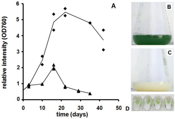
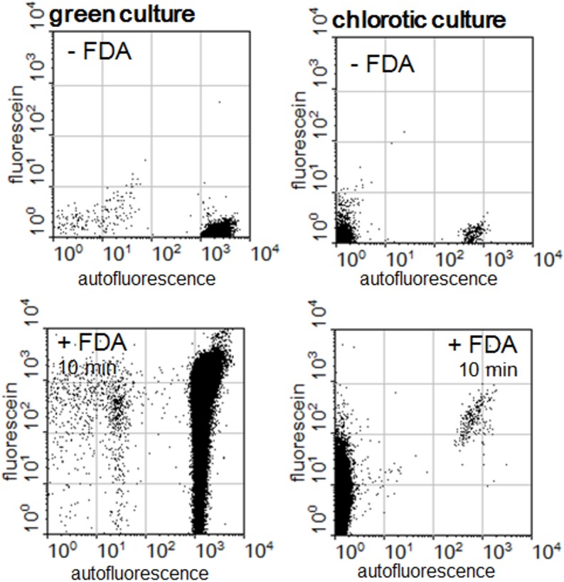
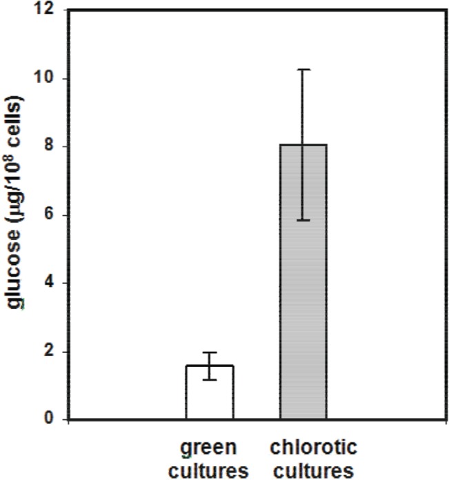
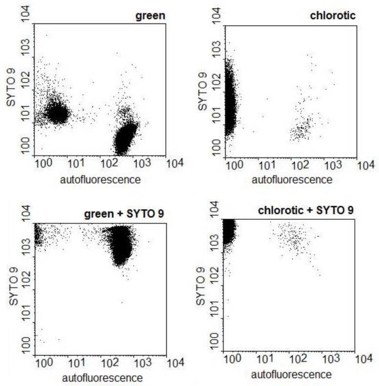
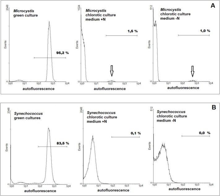
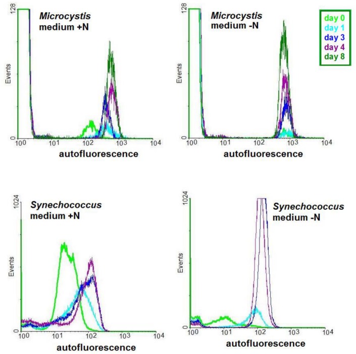
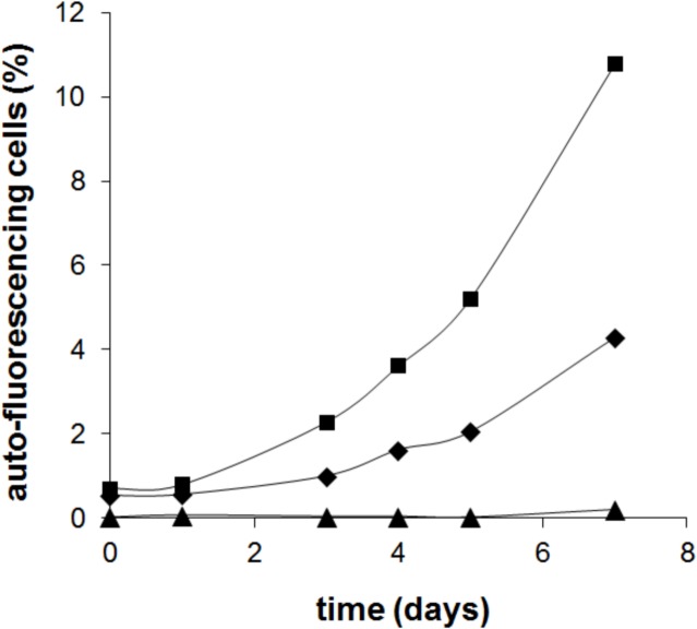
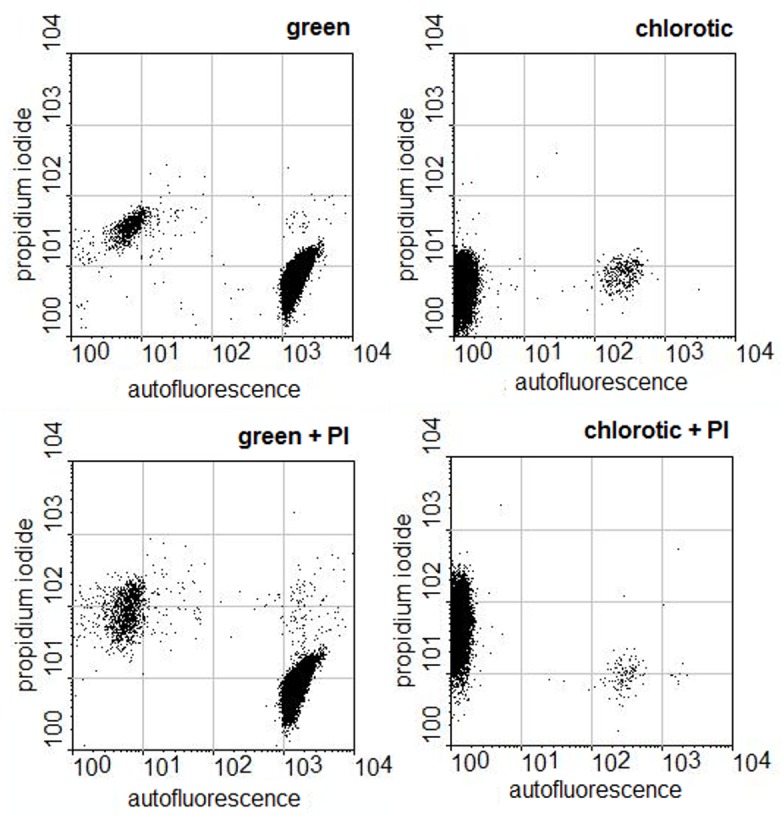
Cultures from the cyanobacterial strain Microcystis aeruginosa PCC 7806 submitted to nutrient limitation become chlorotic. When returned to nutrient rich conditions these cultures regain their green colour. The aim of this study was to verify whether the cells in these cultures could be considered resting stages allowing the survival of periods of nutrient starvation as has been reported for Synechococcus PCC 7942. The experiments with Microcystis were carried out in parallel with Synechococcus cultures to rule out the possibility that any results obtained with Microcystis were due to our particular experimental conditions. The results of the experiments with Synechococcus PCC 7942 cultures were comparable to the reported in the literature. For Microcystis PCC 7806 a different response was observed. Analysis of chlorotic Microcystis cultures by flow cytometry showed that the phenotype of the cells in the population was not homogenous: the amount of nucleic acids was about the same in all cells but only around one percent of the population emitted red autofluorescence indicating the presence of chlorophyll. Monitoring of the reversion of chlorosis by flow cytometry showed that the re-greening was most likely the result of the division of the small population of red autofluorescent cells originally present in the chlorotic cultures. This assumption was confirmed by analysing the integrity of the DNA and the membrane permeability of the cells of chlorotic cultures. Most of the DNA of these cultures was degraded and only the autofluorescent population of the chlorotic cultures showed membrane integrity. Thus, contrary to what has been reported for other cyanobacterial genera, most of the cells in chlorotic Microcystis cultures are not resting stages but dead. It is interesting to note that the red autofluorescent cells of green and chlorotic cultures obtained in double strength ASM-1 medium differ with respect to metabolism: levels of emission of red autofluorescence are higher in the cells of green cultures and the ability to convert fluorescein diacetate of these cells are heterogeneous when compared to the autofluorescent cells of chlorotic cultures. Thus, the small population of the red autofluorescent cells of chlorotic cultures are in a differentiated metabolic state that allow them to persist in conditions in which most of the population loses viability; persistent cells can be detected in chlorotic cultures maintained for more than a year.
对铜绿微囊藻PCC 7806菌株进行营养限制培养时,藻体会变为黄绿色。当恢复到营养丰富的条件时,这些培养物会重新变回绿色。本研究的目的是验证这些培养物中的细胞是否可被视为静止期,从而能够在营养饥饿时期存活下来,就像聚球藻PCC 7942所报道的那样。对微囊藻进行的实验与聚球藻培养物平行进行,以排除微囊藻所获得的任何结果是由于我们特定实验条件所致的可能性。聚球藻PCC 7942培养物的实验结果与文献报道相当。对于微囊藻PCC 7806,观察到了不同的反应。通过流式细胞术对黄绿色微囊藻培养物进行分析表明,群体中细胞的表型并不均一:所有细胞中的核酸量大致相同,但只有约1%的群体发出红色自发荧光,表明存在叶绿素。通过流式细胞术监测黄化逆转过程表明,重新变绿很可能是最初存在于黄化培养物中的少量红色自发荧光细胞分裂的结果。通过分析黄化培养物细胞的DNA完整性和膜通透性,证实了这一假设。这些培养物中的大部分DNA已降解,只有黄化培养物的自发荧光群体显示出膜完整性。因此,与其他蓝藻属所报道的情况相反,黄化微囊藻培养物中的大多数细胞不是静止期,而是死亡的。有趣的是,在双倍强度的ASM-1培养基中获得的绿色和黄化培养物的红色自发荧光细胞在代谢方面存在差异:绿色培养物细胞中的红色自发荧光发射水平较高,与黄化培养物的自发荧光细胞相比,这些细胞转化二乙酸荧光素的能力是异质的。因此,黄化培养物中少量的红色自发荧光细胞处于分化的代谢状态,这使它们能够在大多数群体失去活力的条件下存活;在维持一年以上的黄化培养物中可以检测到持久细胞。