• 文献检索
  • 文档翻译
  • 深度研究
  • 学术资讯
  • Suppr Zotero 插件Zotero 插件
  • 邀请有礼
  • 套餐&价格
  • 历史记录
应用&插件
Suppr Zotero 插件Zotero 插件浏览器插件Mac 客户端Windows 客户端微信小程序
定价
高级版会员购买积分包购买API积分包
服务
文献检索文档翻译深度研究API 文档MCP 服务
关于我们
关于 Suppr公司介绍联系我们用户协议隐私条款
关注我们

Suppr 超能文献

核心技术专利:CN118964589B侵权必究
粤ICP备2023148730 号-1Suppr @ 2026

文献检索

告别复杂PubMed语法,用中文像聊天一样搜索,搜遍4000万医学文献。AI智能推荐,让科研检索更轻松。

立即免费搜索

文件翻译

保留排版,准确专业,支持PDF/Word/PPT等文件格式,支持 12+语言互译。

免费翻译文档

深度研究

AI帮你快速写综述,25分钟生成高质量综述,智能提取关键信息,辅助科研写作。

立即免费体验

非缺血性心肌病的磁共振成像:图文综述

Magnetic Resonance Imaging of Non-ischemic Cardiomyopathies: A Pictorial Essay.

作者信息

Olivas-Chacon Cristina I, Mullins Carola, Stewart Kevan, Akle Nassim, Calleros Jesus E, Ramos-Duran Luis R

机构信息

Department of Radiology, Texas Tech University Health Science Center El Paso, El Paso, Texas, USA.

出版信息

J Clin Imaging Sci. 2015 Jun 30;5:37. doi: 10.4103/2156-7514.159564. eCollection 2015.

DOI:10.4103/2156-7514.159564
PMID:26199786
原文链接:https://pmc.ncbi.nlm.nih.gov/articles/PMC4498316/
Abstract

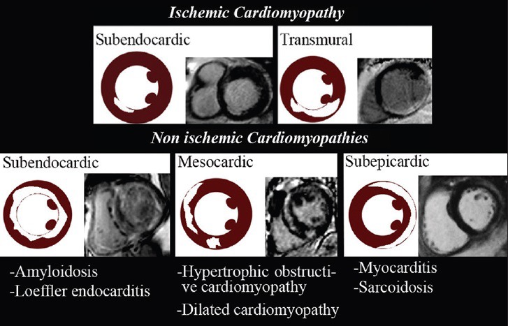
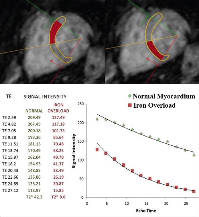
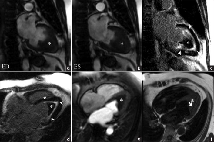
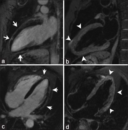
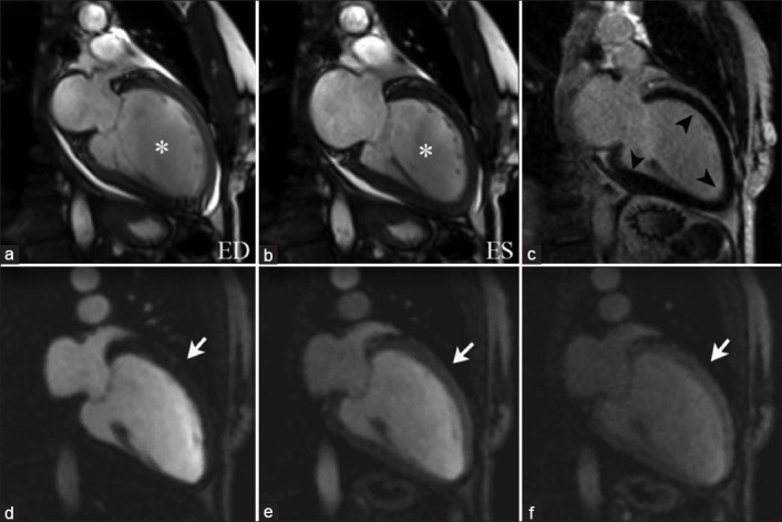
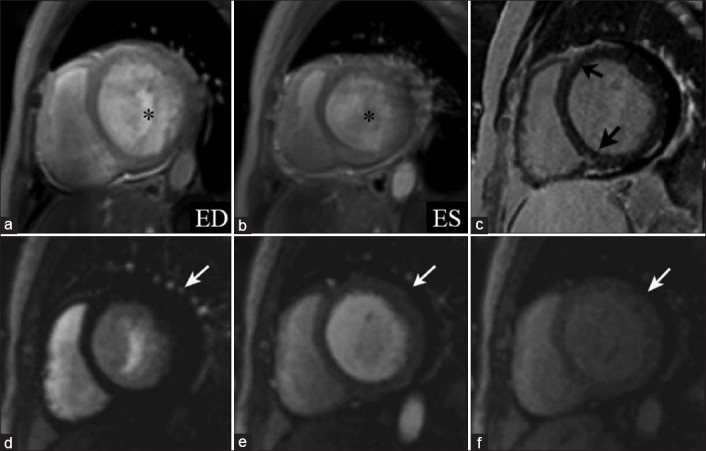
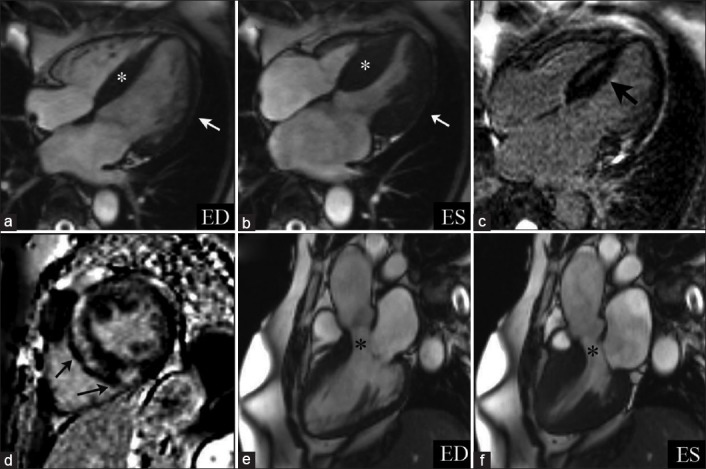
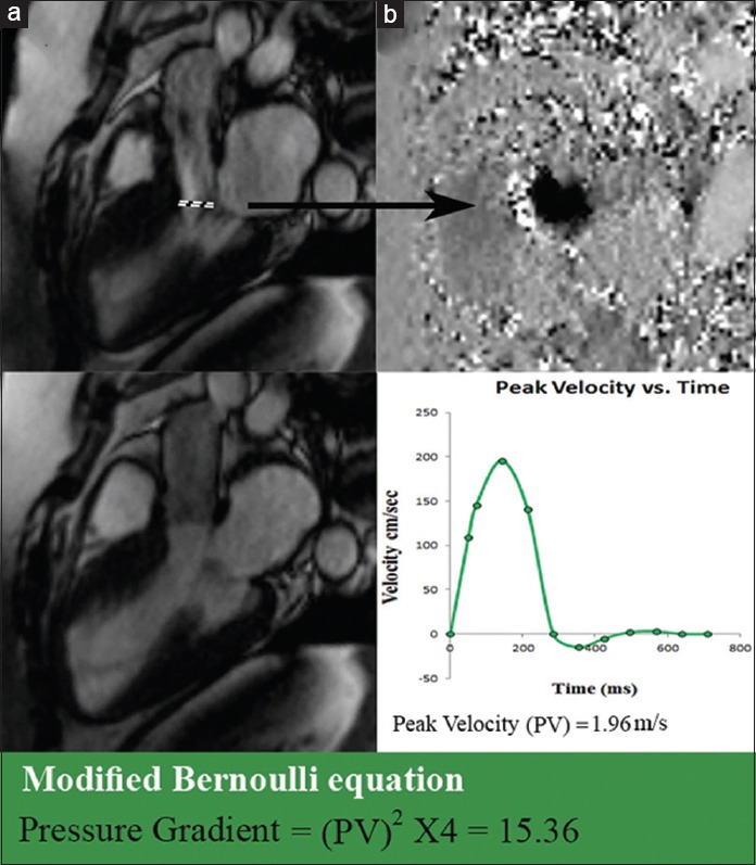
Non-ischemic cardiomyopathies are defined as either primary or secondary diseases of the myocardium resulting in cardiac dysfunction. While primary cardiomyopathies are confined to the heart and can be genetic or acquired, secondary cardiomyopathies show involvement of the heart as a manifestation of an underlying systemic disease including metabolic, inflammatory, granulomatous, infectious, or autoimmune entities. Non-ischemic cardiomyopathies are currently classified as hypertrophic, dilated, restrictive, or unclassifiable, including left ventricular non-compaction. Cardiovascular Magnetic Resonance Imaging (CMRI) not only has the capability to assess cardiac morphology and function, but also the ability to detect edema, hemorrhage, fibrosis, and intramyocardial deposits, providing a valuable imaging tool in the characterization of non-ischemic cardiomyopathies. This pictorial essay shows some of the most important non-ischemic cardiomyopathies with an emphasis on magnetic resonance imaging features.

摘要

非缺血性心肌病被定义为导致心脏功能障碍的原发性或继发性心肌疾病。原发性心肌病局限于心脏,可为遗传性或后天获得性,而继发性心肌病则表现为心脏受累,是潜在全身性疾病的一种表现,包括代谢性、炎症性、肉芽肿性、感染性或自身免疫性疾病。非缺血性心肌病目前分为肥厚型、扩张型、限制型或无法分类型,包括左心室心肌致密化不全。心血管磁共振成像(CMRI)不仅能够评估心脏形态和功能,还能够检测水肿、出血、纤维化和心肌内沉积物,为非缺血性心肌病的特征描述提供了一种有价值的成像工具。这篇图文并茂的文章展示了一些最重要的非缺血性心肌病,并重点介绍了磁共振成像特征。

https://cdn.ncbi.nlm.nih.gov/pmc/blobs/490b/4498316/6276eacd17c5/JCIS-5-37-g022.jpg
https://cdn.ncbi.nlm.nih.gov/pmc/blobs/490b/4498316/9ce143481dae/JCIS-5-37-g003.jpg
https://cdn.ncbi.nlm.nih.gov/pmc/blobs/490b/4498316/19befa36e74e/JCIS-5-37-g005.jpg
https://cdn.ncbi.nlm.nih.gov/pmc/blobs/490b/4498316/2d6327db7156/JCIS-5-37-g007.jpg
https://cdn.ncbi.nlm.nih.gov/pmc/blobs/490b/4498316/1945bb468bf7/JCIS-5-37-g009.jpg
https://cdn.ncbi.nlm.nih.gov/pmc/blobs/490b/4498316/74e6449fbf3b/JCIS-5-37-g011.jpg
https://cdn.ncbi.nlm.nih.gov/pmc/blobs/490b/4498316/eac8cd09d8f9/JCIS-5-37-g012.jpg
https://cdn.ncbi.nlm.nih.gov/pmc/blobs/490b/4498316/4866c99e44a8/JCIS-5-37-g014.jpg
https://cdn.ncbi.nlm.nih.gov/pmc/blobs/490b/4498316/500881016fbc/JCIS-5-37-g015.jpg
https://cdn.ncbi.nlm.nih.gov/pmc/blobs/490b/4498316/e3b2fd7b06bd/JCIS-5-37-g018.jpg
https://cdn.ncbi.nlm.nih.gov/pmc/blobs/490b/4498316/c8a12873b2ff/JCIS-5-37-g019.jpg
https://cdn.ncbi.nlm.nih.gov/pmc/blobs/490b/4498316/32a2e985bff3/JCIS-5-37-g021.jpg
https://cdn.ncbi.nlm.nih.gov/pmc/blobs/490b/4498316/6276eacd17c5/JCIS-5-37-g022.jpg

相似文献

1
Magnetic Resonance Imaging of Non-ischemic Cardiomyopathies: A Pictorial Essay.非缺血性心肌病的磁共振成像:图文综述
J Clin Imaging Sci. 2015 Jun 30;5:37. doi: 10.4103/2156-7514.159564. eCollection 2015.
2
[Patterns of delayed-enhancement in MRI of ischemic and non-ischemic cardiomyopathies].[缺血性和非缺血性心肌病的MRI延迟强化模式]
Rofo. 2007 Jan;179(1):21-30. doi: 10.1055/s-2006-927204.
3
Measurements in Pediatric Patients with Cardiomyopathies: Comparison of Cardiac Magnetic Resonance Imaging and Echocardiography.小儿心肌病患者的测量:心脏磁共振成像与超声心动图的比较
Cardiology. 2015;131(4):245-50. doi: 10.1159/000381418. Epub 2015 May 12.
4
Assessment of Ischemic Cardiomyopathy Using Cardiovascular Magnetic Resonance Imaging: A Pictorial Review.使用心血管磁共振成像评估缺血性心肌病:图文综述
J Clin Imaging Sci. 2015 May 22;5:28. doi: 10.4103/2156-7514.157604. eCollection 2015.
5
Magnetic resonance imaging of primary cardiomyopathies.原发性心肌病的磁共振成像
J Comput Assist Tomogr. 2003 Sep-Oct;27(5):724-34. doi: 10.1097/00004728-200309000-00009.
6
Contrast-enhanced cardiovascular magnetic resonance in primary and ischemic dilated cardiomyopathy.原发性和缺血性扩张型心肌病的对比增强心血管磁共振成像
J Cardiovasc Med (Hagerstown). 2007 Oct;8(10):821-9. doi: 10.2459/JCM.0b013e3280101e3c.
7
Advanced tissue characterization in non-ischemic cardiomyopathies using contrast-enhanced cardiac magnetic resonance imaging.使用对比增强心脏磁共振成像对非缺血性心肌病进行高级组织特征分析。
Saudi Med J. 2010 Feb;31(2):115-22.
8
MRI in the assessment of non ischemic myocardial diseases.MRI 在非缺血性心肌疾病评估中的应用。
Eur J Radiol. 2012 Jul;81(7):1546-8. doi: 10.1016/j.ejrad.2011.02.012. Epub 2011 Mar 10.
9
Cardiovascular magnetic resonance detects silent heart disease missed by echocardiography in systemic lupus erythematosus.心血管磁共振成像可检测出系统性红斑狼疮患者中超声心动图漏诊的无症状心脏病。
Lupus. 2018 Apr;27(4):564-571. doi: 10.1177/0961203317731533. Epub 2017 Sep 19.
10
Three-dimensional phase-sensitive inversion recovery sequencing in the evaluation of left ventricular myocardial scars in ischemic and non-ischemic cardiomyopathy: comparison to three-dimensional inversion recovery sequencing.三维相敏反转恢复序列在评估缺血性和非缺血性心肌病左心室心肌瘢痕中的应用:与三维反转恢复序列的比较
Eur J Radiol. 2014 Dec;83(12):2159-2166. doi: 10.1016/j.ejrad.2014.09.014. Epub 2014 Sep 30.

引用本文的文献

1
Overlapping Phenotypes and Degree of Ventricular Dilatation Are Associated with Severity of Systolic Impairment and Late Gadolinium Enhancement in Non-Ischemic Cardiomyopathies.非缺血性心肌病中,重叠表型和心室扩张程度与收缩功能障碍的严重程度和晚期钆增强有关。
Med Sci Monit. 2018 Jul 22;24:5084-5092. doi: 10.12659/MSM.909172.
2
Supplementary Diagnostic Landmarks of Left Ventricular Non-Compaction on Magnetic Resonance Imaging.磁共振成像上左心室心肌致密化不全的补充诊断标志
Yonsei Med J. 2018 Jan;59(1):63-71. doi: 10.3349/ymj.2018.59.1.63.

本文引用的文献

1
Value of CMR for the differential diagnosis of cardiac masses.CMR 在心脏肿块鉴别诊断中的价值。
JACC Cardiovasc Imaging. 2014 Sep;7(9):896-905. doi: 10.1016/j.jcmg.2014.05.009. Epub 2014 Aug 13.
2
Hypertrophic cardiomyopathy: present and future, with translation into contemporary cardiovascular medicine.肥厚型心肌病:现状与未来,融入当代心血管医学。
J Am Coll Cardiol. 2014 Jul 8;64(1):83-99. doi: 10.1016/j.jacc.2014.05.003.
3
Role of cardiac MRI in the assessment of nonischemic cardiomyopathies.心脏磁共振成像在非缺血性心肌病评估中的作用。
Methodist Debakey Cardiovasc J. 2013 Jul-Sep;9(3):149-55. doi: 10.14797/mdcj-9-3-149.
4
Cardiovascular magnetic resonance artefacts.心血管磁共振伪影。
J Cardiovasc Magn Reson. 2013 May 22;15(1):41. doi: 10.1186/1532-429X-15-41.
5
Multiple left ventricular thrombi in a patient with left ventricular noncompaction.左心室心肌致密化不全患者出现多个左心室血栓。
Tex Heart Inst J. 2012;39(4):550-3.
6
Value of cardiovascular MR in diagnosing left ventricular non-compaction cardiomyopathy and in discriminating between other cardiomyopathies.心血管磁共振在诊断左心室心肌致密化不全心肌病和鉴别其他心肌病中的价值。
Eur Radiol. 2012 Dec;22(12):2699-709. doi: 10.1007/s00330-012-2554-7. Epub 2012 Jul 10.
7
Assessment of left ventricular outflow gradient: hypertrophic cardiomyopathy versus aortic valvular stenosis.评估左心室流出道梯度:肥厚型心肌病与主动脉瓣狭窄。
JACC Cardiovasc Interv. 2012 Jun;5(6):675-81. doi: 10.1016/j.jcin.2012.01.026.
8
Pericardial delayed hyperenhancement with CMR imaging in patients with constrictive pericarditis undergoing surgical pericardiectomy: a case series with histopathological correlation.CMR 成像显示缩窄性心包炎患者心包延迟强化:外科心包切除术病例系列及与组织病理学相关性。
JACC Cardiovasc Imaging. 2011 Nov;4(11):1180-91. doi: 10.1016/j.jcmg.2011.08.011.
9
Cardiac magnetic resonance of left ventricular trabeculation: the new normal.左心室小梁的心脏磁共振成像:新的正常表现
Circ Cardiovasc Imaging. 2011 Mar;4(2):84-6. doi: 10.1161/CIRCIMAGING.110.962472.
10
Regional thicknesses and thickening of compacted and trabeculated myocardial layers of the normal left ventricle studied by cardiovascular magnetic resonance.心血管磁共振研究正常左心室致密心肌层和小梁心肌层的局部厚度和增厚。
Circ Cardiovasc Imaging. 2011 Mar;4(2):139-46. doi: 10.1161/CIRCIMAGING.110.960229. Epub 2010 Dec 30.