• 文献检索
  • 文档翻译
  • 深度研究
  • 学术资讯
  • Suppr Zotero 插件Zotero 插件
  • 邀请有礼
  • 套餐&价格
  • 历史记录
应用&插件
Suppr Zotero 插件Zotero 插件浏览器插件Mac 客户端Windows 客户端微信小程序
定价
高级版会员购买积分包购买API积分包
服务
文献检索文档翻译深度研究API 文档MCP 服务
关于我们
关于 Suppr公司介绍联系我们用户协议隐私条款
关注我们

Suppr 超能文献

核心技术专利:CN118964589B侵权必究
粤ICP备2023148730 号-1Suppr @ 2026

文献检索

告别复杂PubMed语法,用中文像聊天一样搜索,搜遍4000万医学文献。AI智能推荐,让科研检索更轻松。

立即免费搜索

文件翻译

保留排版,准确专业,支持PDF/Word/PPT等文件格式,支持 12+语言互译。

免费翻译文档

深度研究

AI帮你快速写综述,25分钟生成高质量综述,智能提取关键信息,辅助科研写作。

立即免费体验

通过袖珍成像设备检测经常吸烟者右心受累的早期标志物。

Early markers of right heart involvement in regular smokers by Pocket Size Imaging Device.

作者信息

Schiano-Lomoriello Vincenzo, Esposito Roberta, Santoro Ciro, de Simone Giovanni, Galderisi Maurizio

机构信息

Hypertension Research Center (CIRIAPA), Federico II University Hospital, Naples, Italy.

Department of Translational Medical Sciences, Federico II University Hospital, Naples, Italy.

出版信息

Cardiovasc Ultrasound. 2015 Jul 23;13:33. doi: 10.1186/s12947-015-0024-5.

DOI:10.1186/s12947-015-0024-5
PMID:26201963
原文链接:https://pmc.ncbi.nlm.nih.gov/articles/PMC4511232/
Abstract

PURPOSE

To test the diagnostic power of Pocket Size Imaging Device (PSID) in detecting early signs of right heart (RH) involvement in regular smokers (RS) free of overt cardiac involvement.

METHODS

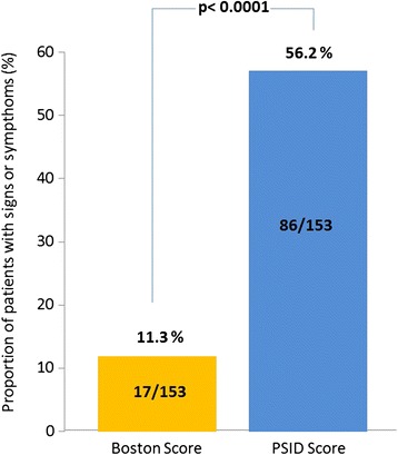
One-hundred-forty-three regular smokers and 51 healthy controls, comparable for age and sex, underwent physical exam (PE), PSID exam and standard echocardiography. Based on a simplified Boston score, ≥1 of clinical signs (jugular venous distension, hepatomegaly, peripheral pitting oedema and abnormal pulmonary sounds) were considered indicative of RH involvement. A composite score (1 to 4) obtained by summing the points of four quantitative RH abnormalities detectable by PSID (inferior vena cava [IVC] dilatation, reduced IVC respiratory variation, right ventricular dilatation and right atrial dilatation), was generated and ≥1 of PSID abnormal signs was considered indicative of RH involvement.

RESULTS

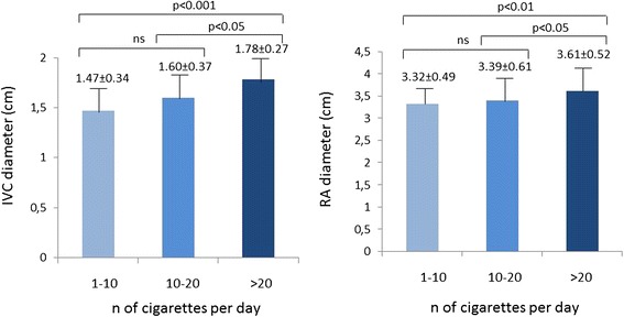
Boston score was not significantly different between the two groups. By using PSID, smokers exhibited greater IVC diameter (p < 0.0001), right atrial diameter (p < 0.002) and higher PSID score (p < 0.005) than controls. Compared to PE, the additional diagnostic power of PSID (≥1 abnormal sign of both Boston and PSID score) was 44.9% in smokers. By dividing smokers in tertiles according to number of cigarettes per day, the third tertile showed the largest values of both IVC and right atrial dimension. Differences were confirmed by standard echocardiography. Reproducibility of PSID measurements and concordance of linear measurements between PSID and standard echo measurements was very good except for concordance of right ventricular basal diameter.

CONCLUSIONS

PSID detects early ultrasound signs of RH involvement in regular otherwise healthy smokers in comparison with PE.

摘要

目的

测试袖珍成像设备(PSID)在检测无明显心脏受累的规律吸烟者(RS)右心(RH)受累早期迹象方面的诊断能力。

方法

143名规律吸烟者和51名年龄和性别匹配的健康对照者接受了体格检查(PE)、PSID检查和标准超声心动图检查。根据简化的波士顿评分,临床体征(颈静脉扩张、肝肿大、外周凹陷性水肿和异常肺部啰音)中≥1项被认为提示RH受累。通过将PSID可检测到的四项定量RH异常(下腔静脉[IVC]扩张、IVC呼吸变化减小、右心室扩张和右心房扩张)的分数相加得到一个综合评分(1至4分),PSID异常体征中≥1项被认为提示RH受累。

结果

两组之间的波士顿评分无显著差异。使用PSID时,吸烟者的IVC直径(p<0.0001)、右心房直径(p<0.002)和PSID评分更高(p<0.005)。与PE相比,PSID(波士顿和PSID评分均≥1项异常体征)在吸烟者中的额外诊断能力为44.9%。根据每日吸烟量将吸烟者分为三分位数,第三三分位数的IVC和右心房尺寸值最大。标准超声心动图证实了差异。PSID测量的可重复性以及PSID与标准回声测量之间线性测量的一致性非常好,除了右心室基底直径的一致性。

结论

与PE相比,PSID可检测到无其他明显疾病的规律吸烟者RH受累的早期超声迹象。

https://cdn.ncbi.nlm.nih.gov/pmc/blobs/6d62/4511232/f445ae0d80c4/12947_2015_24_Fig2_HTML.jpg
https://cdn.ncbi.nlm.nih.gov/pmc/blobs/6d62/4511232/dfe2b05d02f8/12947_2015_24_Fig1_HTML.jpg
https://cdn.ncbi.nlm.nih.gov/pmc/blobs/6d62/4511232/f445ae0d80c4/12947_2015_24_Fig2_HTML.jpg
https://cdn.ncbi.nlm.nih.gov/pmc/blobs/6d62/4511232/dfe2b05d02f8/12947_2015_24_Fig1_HTML.jpg
https://cdn.ncbi.nlm.nih.gov/pmc/blobs/6d62/4511232/f445ae0d80c4/12947_2015_24_Fig2_HTML.jpg

相似文献

1
Early markers of right heart involvement in regular smokers by Pocket Size Imaging Device.通过袖珍成像设备检测经常吸烟者右心受累的早期标志物。
Cardiovasc Ultrasound. 2015 Jul 23;13:33. doi: 10.1186/s12947-015-0024-5.
2
Pocket-size imaging device: effectiveness for ward-based transthoracic studies.口袋大小的成像设备:在病房进行经胸研究的有效性。
Eur Heart J Cardiovasc Imaging. 2013 Dec;14(12):1132-9. doi: 10.1093/ehjci/jet091. Epub 2013 May 24.
3
Identification of the main determinants of abdominal aorta size: a screening by Pocket Size Imaging Device.腹主动脉大小主要决定因素的识别:使用袖珍成像设备进行筛查。
Cardiovasc Ultrasound. 2017 Jan 13;15(1):2. doi: 10.1186/s12947-016-0094-z.
4
Bedside screening with the use of pocket-size imaging device can be useful for ruling out carotid artery stenosis in patients scheduled for cardiac surgery.使用袖珍成像设备进行床边筛查,对于排除计划进行心脏手术患者的颈动脉狭窄可能有用。
Echocardiography. 2017 May;34(5):716-722. doi: 10.1111/echo.13507. Epub 2017 Mar 16.
5
Improved cardiovascular diagnostic accuracy by pocket size imaging device in non-cardiologic outpatients: the NaUSiCa (Naples Ultrasound Stethoscope in Cardiology) study.口袋大小的成像设备提高非心脏病门诊患者的心血管诊断准确性:NaUSiCa(那不勒斯心脏病学超声听诊器)研究
Cardiovasc Ultrasound. 2010 Nov 26;8:51. doi: 10.1186/1476-7120-8-51.
6
Three-dimensional echocardiography and 2D-3D speckle-tracking imaging in chronic pulmonary hypertension: diagnostic accuracy in detecting hemodynamic signs of right ventricular (RV) failure.三维超声心动图和二维-三维斑点追踪成像在慢性肺动脉高压中的应用:检测右心室(RV)衰竭血流动力学征象的诊断准确性
J Am Heart Assoc. 2015 Mar 19;4(3):e001584. doi: 10.1161/JAHA.114.001584.
7
Systemic venous diameters, collapsibility indices, and right atrial measurements in normal pediatric subjects.正常儿科受检者的体循环静脉直径、塌陷指数和右心房测量值。
J Am Soc Echocardiogr. 2014 Feb;27(2):155-62. doi: 10.1016/j.echo.2013.09.002. Epub 2013 Oct 9.
8
Personal mobile device-based pocket echocardiograph-The diagnostic value and clinical utility.基于个人移动设备的掌上超声心动图——诊断价值和临床应用。
Adv Med Sci. 2019 Mar;64(1):157-161. doi: 10.1016/j.advms.2018.11.003. Epub 2019 Jan 25.
9
Estimation of right ventricular dysfunction by computed tomography pulmonary angiography: a valuable adjunct for evaluating the severity of acute pulmonary embolism.通过计算机断层扫描肺动脉造影评估右心室功能障碍:评估急性肺栓塞严重程度的一项有价值的辅助手段。
J Thromb Thrombolysis. 2017 Feb;43(2):271-278. doi: 10.1007/s11239-016-1438-0.
10
Brief cardiovascular imaging with pocket-size ultrasound devices improves the accuracy of the initial assessment of suspected pulmonary embolism.使用口袋大小的超声设备进行简要心血管成像可提高疑似肺栓塞初始评估的准确性。
Int J Cardiovasc Imaging. 2018 Oct;34(10):1595-1605. doi: 10.1007/s10554-018-1382-5. Epub 2018 May 30.

引用本文的文献

1
Focused cardiac ultrasound as a predictor of readmission in acute decompensated heart failure.聚焦心脏超声作为急性失代偿性心力衰竭再入院的预测指标。
Int J Cardiovasc Imaging. 2018 Jul;34(7):1075-1079. doi: 10.1007/s10554-018-1317-1. Epub 2018 Feb 15.
2
Results of ACCORDIAN in ACCORD with lower blood pressure begetting lower mortality in patients with diabetes.ACCORDIAN 研究显示,糖尿病患者的降压幅度与死亡率降低相关。
Diabetes Obes Metab. 2018 Jun;20(6):1335-1336. doi: 10.1111/dom.13249. Epub 2018 Feb 28.
3
Identification of the main determinants of abdominal aorta size: a screening by Pocket Size Imaging Device.

本文引用的文献

1
Recommendations for cardiac chamber quantification by echocardiography in adults: an update from the American Society of Echocardiography and the European Association of Cardiovascular Imaging.成人经超声心动图进行心腔定量的建议:美国超声心动图学会和欧洲心血管影像学会的更新版
Eur Heart J Cardiovasc Imaging. 2015 Mar;16(3):233-70. doi: 10.1093/ehjci/jev014.
2
Adverse effects of cigarette and noncigarette smoke exposure on the autonomic nervous system: mechanisms and implications for cardiovascular risk.香烟和非香烟烟雾暴露对自主神经系统的不良影响:对心血管风险的机制和意义。
J Am Coll Cardiol. 2014 Oct 21;64(16):1740-50. doi: 10.1016/j.jacc.2014.06.1201.
3
腹主动脉大小主要决定因素的识别:使用袖珍成像设备进行筛查。
Cardiovasc Ultrasound. 2017 Jan 13;15(1):2. doi: 10.1186/s12947-016-0094-z.
Echocardiographic assessment of estimated right atrial pressure and size predicts mortality in pulmonary arterial hypertension.
经超声心动图评估估计的右心房压力和大小可预测肺动脉高压患者的死亡率。
Chest. 2015 Jan;147(1):198-208. doi: 10.1378/chest.13-3035.
4
Smoking and cardiovascular disease: mechanisms of endothelial dysfunction and early atherogenesis.吸烟与心血管疾病:内皮功能障碍与早期动脉粥样硬化形成的机制。
Arterioscler Thromb Vasc Biol. 2014 Mar;34(3):509-15. doi: 10.1161/ATVBAHA.113.300156.
5
Pocket-size imaging device: effectiveness for ward-based transthoracic studies.口袋大小的成像设备:在病房进行经胸研究的有效性。
Eur Heart J Cardiovasc Imaging. 2013 Dec;14(12):1132-9. doi: 10.1093/ehjci/jet091. Epub 2013 May 24.
6
Incremental value of pocket-sized imaging device for bedside diagnosis of unilateral pleural effusions and ultrasound-guided thoracentesis.袖珍成像设备在单侧胸腔积液床边诊断及超声引导下胸腔穿刺术中的增量价值
Interact Cardiovasc Thorac Surg. 2012 Oct;15(4):596-601. doi: 10.1093/icvts/ivs223. Epub 2012 Jul 19.
7
Diagnostic performance of handheld echocardiography for the assessment of basic cardiac morphology and function: a validation study in routine cardiac patients.手持式超声心动图评估心脏基本形态和功能的诊断性能:一项针对常规心脏病患者的验证研究。
Echocardiography. 2012 Sep;29(8):887-94. doi: 10.1111/j.1540-8175.2012.01728.x. Epub 2012 May 29.
8
Fast track echo of abdominal aortic aneurysm using a real pocket-ultrasound device at bedside.使用床边便携式超声设备对腹主动脉瘤进行快速超声检查。
Echocardiography. 2012 Mar;29(3):285-90. doi: 10.1111/j.1540-8175.2011.01559.x. Epub 2011 Nov 8.
9
Diagnostic utility and clinical usefulness of the pocket echocardiographic device.便携式超声心动图设备的诊断效用及临床应用价值
Echocardiography. 2012;29(1):1-6. doi: 10.1111/j.1540-8175.2011.01553.x. Epub 2011 Nov 2.
10
Methodological approach for the assessment of ultrasound reproducibility of cardiac structure and function: a proposal of the study group of Echocardiography of the Italian Society of Cardiology (Ultra Cardia SIC) part I.心脏结构和功能超声可重复性评估的方法学探讨:意大利心脏病学会超声心动图研究组(Ultra Cardia SIC)提案 第一部分
Cardiovasc Ultrasound. 2011 Sep 26;9:26. doi: 10.1186/1476-7120-9-26.