Esposito Roberta, Ilardi Federica, Schiano Lomoriello Vincenzo, Sorrentino Regina, Sellitto Vincenzo, Giugliano Giuseppe, Esposito Giovanni, Trimarco Bruno, Galderisi Maurizio
Department of Advanced Biomedical Sciences, Division of Cardiology, Federico II University Hospital, Naples, Italy.
Interdepartimental Laboratory of Cardiac Imaging, Federico II University Hospital, Via S. Pansini 5,bld 1, 80131, Naples, Italy.
Cardiovasc Ultrasound. 2017 Jan 13;15(1):2. doi: 10.1186/s12947-016-0094-z.
Ultrasound exam as a screening test for abdominal aorta (AA) can visualize the aorta in 99% of patients and has a sensitivity and specificity approaching 100% in screening settings for aortic aneurysm. Pocket Size Imaging Device (PSID) has a potential value as a screening tool, because of its possible use in several clinical settings. Our aim was to assess the impact of demographics and cardiovascular (CV) risk factors on AA size by using PSID in an outpatient screening.
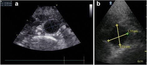
Consecutive patients, referring for a CV assessment in a 6 months period, were screened. AA was visualized by subcostal view in longitudinal and transverse plans in order to determine the greatest anterior-posterior diameter. After excluding 5 patients with AA aneurysm, 508 outpatients were enrolled. All patients underwent a sequential assessment including clinical history with collection of CV risk factors, physical examination, PSID exam and standard Doppler echoc exam using a 2.5 transducer with harmonic capability, both by expert ultrasound operators, during the same morning. Standard echocardiography operators were blinded on PSID exam and viceversa.
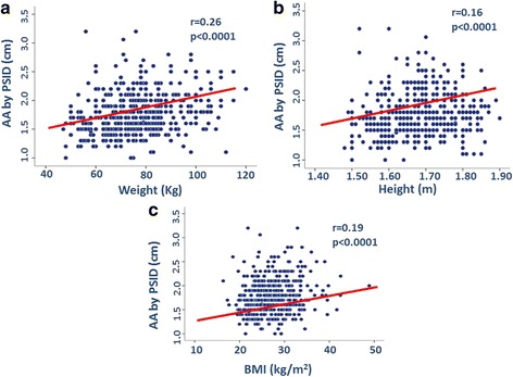
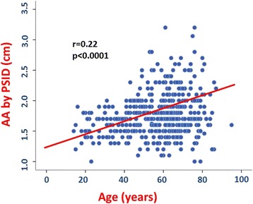
Diagnostic accuracy of AA size by PSID was tested successfully with standard echo machine in a subgroup (n = 102) (rho = 0.966, p < 0.0001). AA diameter was larger in men than in women and in ≥50 -years old subjects than in those <50 -years old (both p < 0.0001). AA was larger in patients with coronary artery disease (CAD) (p < 0.0001). By a multivariate model, male sex (p < 0.0001), age and body mass index (both p < 0.0001), CAD (p < 0.01) and heart rate (p = 0.018) were independent predictors of AA size (cumulative R = 0.184, p < 0.0001).
PSID is a reliable tool for the screening of determinants of AA size. AA diameter is greater in men and strongly influenced by aging and overweight. CAD may be also associated to increased AA diameter.
超声检查作为腹主动脉(AA)的筛查试验,在99%的患者中能够显示主动脉,并且在主动脉瘤筛查中其敏感性和特异性接近100%。袖珍成像设备(PSID)作为一种筛查工具具有潜在价值,因为它可能适用于多种临床场景。我们的目的是通过在门诊筛查中使用PSID来评估人口统计学和心血管(CV)危险因素对AA大小的影响。
对连续6个月前来进行CV评估的患者进行筛查。通过肋下视图在纵向和横向平面观察AA,以确定最大前后径。排除5例患有AA动脉瘤的患者后,纳入508例门诊患者。所有患者在同一天上午由专业超声操作员进行连续评估,包括收集CV危险因素的临床病史、体格检查、PSID检查以及使用具有谐波功能的2.5探头进行标准多普勒超声心动图检查。标准超声心动图操作员对PSID检查结果不知情,反之亦然。
在一个亚组(n = 102)中,PSID测量AA大小的诊断准确性通过标准超声机器成功得到验证(rho = 0.966,p < 0.0001)。男性的AA直径大于女性,≥50岁受试者的AA直径大于<50岁的受试者(p均< 0.0001)。患有冠状动脉疾病(CAD)的患者AA直径更大(p < 0.0001)。通过多变量模型,男性性别(p < 0.0001)、年龄和体重指数(p均< 0.0001)、CAD(p < 0.01)和心率(p = 0.018)是AA大小的独立预测因素(累积R = 0.184,p < 0.0001)。
PSID是筛查AA大小决定因素的可靠工具。男性的AA直径更大,且受衰老和超重的影响较大。CAD也可能与AA直径增加有关。