• 文献检索
  • 文档翻译
  • 深度研究
  • 学术资讯
  • Suppr Zotero 插件Zotero 插件
  • 邀请有礼
  • 套餐&价格
  • 历史记录
应用&插件
Suppr Zotero 插件Zotero 插件浏览器插件Mac 客户端Windows 客户端微信小程序
定价
高级版会员购买积分包购买API积分包
服务
文献检索文档翻译深度研究API 文档MCP 服务
关于我们
关于 Suppr公司介绍联系我们用户协议隐私条款
关注我们

Suppr 超能文献

核心技术专利:CN118964589B侵权必究
粤ICP备2023148730 号-1Suppr @ 2026

文献检索

告别复杂PubMed语法,用中文像聊天一样搜索,搜遍4000万医学文献。AI智能推荐,让科研检索更轻松。

立即免费搜索

文件翻译

保留排版,准确专业,支持PDF/Word/PPT等文件格式,支持 12+语言互译。

免费翻译文档

深度研究

AI帮你快速写综述,25分钟生成高质量综述,智能提取关键信息,辅助科研写作。

立即免费体验

在亚临床肥厚型心肌病中,室间隔会异常凸向左心室。

Abnormal septal convexity into the left ventricle occurs in subclinical hypertrophic cardiomyopathy.

作者信息

Reant Patricia, Captur Gabriella, Mirabel Mariana, Nasis Arthur, M Sado Daniel, Maestrini Viviana, Castelletti Silvia, Manisty Charlotte, Herrey Anna S, Syrris Petros, Tome-Esteban Maite, Jenkins Sharon, Elliott Perry M, McKenna William J, Moon James C

机构信息

Division of Cardiovascular Imaging and Inherited Cardiac Disease Unit, The Heart Hospital, Institute of Cardiovascular Science, University College London, London, UK.

Hôpital Cardiologique du Haut-Levêque (Pessac), CHU de Bordeaux, Université de Bordeaux, Bordeaux, France.

出版信息

J Cardiovasc Magn Reson. 2015 Jul 30;17(1):64. doi: 10.1186/s12968-015-0160-y.

DOI:10.1186/s12968-015-0160-y
PMID:26219660
原文链接:https://pmc.ncbi.nlm.nih.gov/articles/PMC4518641/
Abstract

BACKGROUND

Sarcomeric gene mutations cause hypertrophic cardiomyopathy (HCM). In gene mutation carriers without left ventricular (LV) hypertrophy (G + LVH-), subclinical imaging biomarkers are recognized as predictors of overt HCM, consisting of anterior mitral valve leaflet elongation, myocardial crypts, hyperdynamic LV ejection fraction, and abnormal apical trabeculation. Reverse curvature of the interventricular septum (into the LV) is characteristic of overt HCM. We aimed to assess LV septal convexity in subclinical HCM.

METHODS

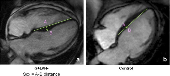
Cardiovascular magnetic resonance was performed on 36 G + LVH- individuals (31 ± 14 years, 33 % males) with a pathogenic sarcomere mutation, and 36 sex and age-matched healthy controls (33 ± 12 years, 33 % males). Septal convexity (SCx) was measured in the apical four chamber view perpendicular to a reference line connecting the mid-septal wall at tricuspid valve insertion level and the apical right ventricular insertion point.

RESULTS

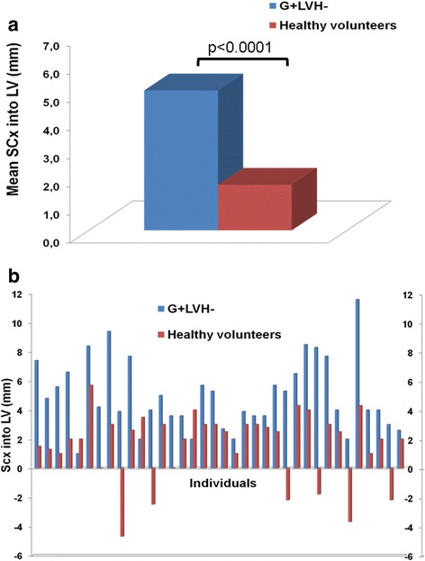
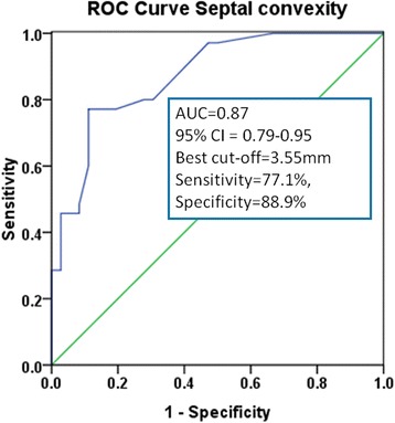
Septal convexity was increased in G + LVH- compared to controls (maximal distance of endocardium to reference line: 5.0 ± 2.5 mm vs. 1.6 ± 2.4 mm, p ≤ 0.0001). Expected findings occurred in G + LVH- individuals: longer anterior mitral valve leaflet (23.5 ± 3.0 mm vs. 19.9 ± 3.1 mm, p ≤ 0.0001), higher relative wall thickness (0.31 ± 0.05 vs. 0.29 ± 0.04, p ≤ 0.05), higher LV ejection fraction (70.8 ± 4.3 % vs. 68.3 ± 4.4 %, p ≤ 0.05), and smaller LV end-systolic volume index (21.4 ± 4.4 ml/m(2) vs. 23.7 ± 5.8 ml/m(2), p ≤ 0.05). Other morphologic measurements (LV angles, sphericity index, and eccentricity index) were not different between G + LVH- and controls.

CONCLUSIONS

Septal convexity is an additional previously undescribed feature of subclinical HCM.

摘要

背景

肌节基因突变可导致肥厚型心肌病(HCM)。在无左心室(LV)肥厚的基因突变携带者(G+LVH-)中,亚临床影像生物标志物被认为是显性HCM的预测指标,包括二尖瓣前叶延长、心肌隐窝、左室射血分数增高及心尖小梁异常。室间隔向LV内反向弯曲是显性HCM的特征。我们旨在评估亚临床HCM中的左室间隔凸度。

方法

对36例携带致病性肌节基因突变的G+LVH-个体(31±14岁,33%为男性)和36例年龄及性别匹配的健康对照者(33±12岁,33%为男性)进行心血管磁共振检查。在垂直于连接三尖瓣附着水平室间隔壁中点与心尖右室附着点的参考线的心尖四腔视图中测量间隔凸度(SCx)。

结果

与对照组相比,G+LVH-个体的间隔凸度增加(心内膜到参考线的最大距离:5.0±2.5mm对1.6±2.4mm,p≤0.0001)。G+LVH-个体出现了预期的表现:二尖瓣前叶更长(23.5±3.0mm对19.9±3.1mm,p≤0.0001)、相对室壁厚度更高(0.31±0.05对0.29±0.04,p≤0.05)、左室射血分数更高(70.8±4.3%对68.3±4.4%,p≤0.05)以及左室收缩末期容积指数更小(21.4±4.4ml/m²对23.7±5.8ml/m²,p≤0.05)。G+LVH-个体与对照组之间的其他形态学测量指标(左室角度、球形指数和偏心指数)无差异。

结论

间隔凸度是亚临床HCM另一个先前未描述的特征。

https://cdn.ncbi.nlm.nih.gov/pmc/blobs/a537/4518641/d16569070231/12968_2015_160_Fig5_HTML.jpg
https://cdn.ncbi.nlm.nih.gov/pmc/blobs/a537/4518641/e961e0665810/12968_2015_160_Fig1_HTML.jpg
https://cdn.ncbi.nlm.nih.gov/pmc/blobs/a537/4518641/efe569e92dc3/12968_2015_160_Fig2_HTML.jpg
https://cdn.ncbi.nlm.nih.gov/pmc/blobs/a537/4518641/303d83eac038/12968_2015_160_Fig3_HTML.jpg
https://cdn.ncbi.nlm.nih.gov/pmc/blobs/a537/4518641/a6a04cca1b72/12968_2015_160_Fig4_HTML.jpg
https://cdn.ncbi.nlm.nih.gov/pmc/blobs/a537/4518641/d16569070231/12968_2015_160_Fig5_HTML.jpg

相似文献

1
Abnormal septal convexity into the left ventricle occurs in subclinical hypertrophic cardiomyopathy.在亚临床肥厚型心肌病中,室间隔会异常凸向左心室。
J Cardiovasc Magn Reson. 2015 Jul 30;17(1):64. doi: 10.1186/s12968-015-0160-y.
2
Prediction of sarcomere mutations in subclinical hypertrophic cardiomyopathy.亚临床肥厚型心肌病肌节突变的预测
Circ Cardiovasc Imaging. 2014 Nov;7(6):863-71. doi: 10.1161/CIRCIMAGING.114.002411. Epub 2014 Sep 16.
3
The Burden of Early Phenotypes and the Influence of Wall Thickness in Hypertrophic Cardiomyopathy Mutation Carriers: Findings From the HCMNet Study.肥厚型心肌病突变携带者的早期表型负担和壁厚度的影响:来自 HCMNet 研究的发现。
JAMA Cardiol. 2017 Apr 1;2(4):419-428. doi: 10.1001/jamacardio.2016.5670.
4
Cardiovascular magnetic resonance of mitral valve length in hypertrophic cardiomyopathy.肥厚型心肌病中二尖瓣长度的心血管磁共振成像
J Cardiovasc Magn Reson. 2016 Jun 4;18(1):33. doi: 10.1186/s12968-016-0250-5.
5
T1 measurements identify extracellular volume expansion in hypertrophic cardiomyopathy sarcomere mutation carriers with and without left ventricular hypertrophy.T1 测量可识别肥厚型心肌病肌节突变携带者有无左心室肥厚的细胞外容积扩张。
Circ Cardiovasc Imaging. 2013 May 1;6(3):415-22. doi: 10.1161/CIRCIMAGING.112.000333. Epub 2013 Apr 2.
6
Genetic relevance and determinants of mitral leaflet size in hypertrophic cardiomyopathy.肥厚型心肌病中二尖瓣叶大小的遗传相关性及决定因素
Cardiovasc Ultrasound. 2019 Oct 28;17(1):21. doi: 10.1186/s12947-019-0171-1.
7
CMR derived left ventricular septal convexity in carriers of the hypertrophic cardiomyopathy-causing MYBPC3-Q1061X mutation.肥厚型心肌病致病基因 MYBPC3-Q1061X 突变携带者的心脏磁共振左室间隔凸面成像。
Sci Rep. 2019 Apr 11;9(1):5960. doi: 10.1038/s41598-019-42376-7.
8
Left Atrial structure and function in hypertrophic cardiomyopathy sarcomere mutation carriers with and without left ventricular hypertrophy.肥厚型心肌病肌节突变携带者左心房结构和功能与左心室肥厚的关系。
J Cardiovasc Magn Reson. 2017 Dec 28;19(1):107. doi: 10.1186/s12968-017-0420-0.
9
Abnormal cardiac formation in hypertrophic cardiomyopathy: fractal analysis of trabeculae and preclinical gene expression.肥厚型心肌病中心脏结构异常:小梁的分形分析及临床前基因表达
Circ Cardiovasc Genet. 2014 Jun;7(3):241-8. doi: 10.1161/CIRCGENETICS.113.000362. Epub 2014 Apr 5.
10
Systolic anterior motion of the anterior mitral valve leaflet begins in subclinical hypertrophic cardiomyopathy.前二尖瓣瓣叶收缩期前向运动始于亚临床肥厚型心肌病。
Eur Heart J Cardiovasc Imaging. 2023 Dec 21;25(1):86-94. doi: 10.1093/ehjci/jead186.

引用本文的文献

1
Left ventricular septal convexity in differentiating hypertrophic cardiomyopathy from hypertensive heart disease - a cardiac magnetic resonance study.通过心脏磁共振成像研究左心室间隔凸度在肥厚型心肌病与高血压性心脏病鉴别诊断中的作用
Sci Rep. 2025 Aug 5;15(1):28499. doi: 10.1038/s41598-025-14357-6.
2
The Role of Cardiovascular Magnetic Resonance Imaging in Athletic Individuals-A Narrative Review.心血管磁共振成像在运动员中的作用——一篇叙述性综述
J Clin Med. 2025 May 20;14(10):3576. doi: 10.3390/jcm14103576.
3
Advancing Cardiovascular Diagnostics: The Expanding Role of CMR in Heart Failure and Cardiomyopathies.

本文引用的文献

1
Abnormal cardiac formation in hypertrophic cardiomyopathy: fractal analysis of trabeculae and preclinical gene expression.肥厚型心肌病中心脏结构异常:小梁的分形分析及临床前基因表达
Circ Cardiovasc Genet. 2014 Jun;7(3):241-8. doi: 10.1161/CIRCGENETICS.113.000362. Epub 2014 Apr 5.
2
Relation between left ventricular outflow tract obstruction and left ventricular shape in patients with hypertrophic cardiomyopathy: a cardiac magnetic resonance imaging study.肥厚型心肌病患者左心室流出道梗阻与左心室形态的关系:一项心脏磁共振成像研究。
Arch Cardiovasc Dis. 2013 Aug-Sep;106(8-9):440-7. doi: 10.1016/j.acvd.2013.05.002. Epub 2013 Jul 29.
3
推进心血管诊断:心脏磁共振成像在心力衰竭和心肌病中不断扩大的作用
J Clin Med. 2025 Jan 28;14(3):865. doi: 10.3390/jcm14030865.
4
Phenotypic Spectrum of Subclinical Sarcomere-Related Hypertrophic Cardiomyopathy and Transition to Overt Disease.临床下肌节相关肥厚型心肌病表型谱及向显性疾病的转变。
Circ Genom Precis Med. 2024 Aug;17(4):e004580. doi: 10.1161/CIRCGEN.124.004580. Epub 2024 Jun 24.
5
Systolic anterior motion of the anterior mitral valve leaflet begins in subclinical hypertrophic cardiomyopathy.前二尖瓣瓣叶收缩期前向运动始于亚临床肥厚型心肌病。
Eur Heart J Cardiovasc Imaging. 2023 Dec 21;25(1):86-94. doi: 10.1093/ehjci/jead186.
6
Role of Imaging in Cardiomyopathies.影像学在心肌病中的作用。
Card Fail Rev. 2023 May 25;9:e08. doi: 10.15420/cfr.2022.26. eCollection 2023.
7
Hypertensive Heart Disease-The Imaging Perspective.高血压性心脏病——影像学视角
J Clin Med. 2023 Apr 25;12(9):3122. doi: 10.3390/jcm12093122.
8
Clinical application of CMR in cardiomyopathies: evolving concepts and techniques : A position paper of myocardial and pericardial diseases and cardiac magnetic resonance working groups of Italian society of cardiology.CMR 在心肌病中的临床应用:不断发展的概念和技术:意大利心脏病学会心肌和心包疾病以及心脏磁共振工作组的立场文件。
Heart Fail Rev. 2023 Jan;28(1):77-95. doi: 10.1007/s10741-022-10235-9. Epub 2022 May 10.
9
Myocardial Perfusion Defects in Hypertrophic Cardiomyopathy Mutation Carriers.肥厚型心肌病突变携带者的心肌灌注缺陷。
J Am Heart Assoc. 2021 Aug 3;10(15):e020227. doi: 10.1161/JAHA.120.020227. Epub 2021 Jul 27.
10
Multi-omics integration identifies key upstream regulators of pathomechanisms in hypertrophic cardiomyopathy due to truncating MYBPC3 mutations.多组学整合鉴定出截断 MYBPC3 突变致肥厚型心肌病病理机制的关键上游调控因子。
Clin Epigenetics. 2021 Mar 23;13(1):61. doi: 10.1186/s13148-021-01043-3.
Deep basal inferoseptal crypts occur more commonly in patients with hypertrophic cardiomyopathy due to disease-causing myofilament mutations.
深层基底间隔隐窝在肥厚型心肌病患者中更为常见,这是由于致病肌丝突变所致。
Radiology. 2013 Oct;269(1):68-76. doi: 10.1148/radiol.13122344. Epub 2013 Jun 14.
4
A systematic review and meta-analysis of genotype-phenotype associations in patients with hypertrophic cardiomyopathy caused by sarcomeric protein mutations.肌节蛋白突变致肥厚型心肌病患者基因型-表型相关性的系统评价和荟萃分析。
Heart. 2013 Dec;99(24):1800-11. doi: 10.1136/heartjnl-2013-303939. Epub 2013 May 14.
5
Relationship between left ventricular sphericity and trabeculation indexes in patients with dilated cardiomyopathy: a cardiac magnetic resonance study.扩张型心肌病患者左心室球形度与小梁化指数的关系:一项心脏磁共振研究。
Eur Heart J Cardiovasc Imaging. 2013 Sep;14(9):914-20. doi: 10.1093/ehjci/jet064. Epub 2013 May 3.
6
T1 measurements identify extracellular volume expansion in hypertrophic cardiomyopathy sarcomere mutation carriers with and without left ventricular hypertrophy.T1 测量可识别肥厚型心肌病肌节突变携带者有无左心室肥厚的细胞外容积扩张。
Circ Cardiovasc Imaging. 2013 May 1;6(3):415-22. doi: 10.1161/CIRCIMAGING.112.000333. Epub 2013 Apr 2.
7
Genetic complexity in hypertrophic cardiomyopathy revealed by high-throughput sequencing.高通量测序揭示肥厚型心肌病的遗传复杂性。
J Med Genet. 2013 Apr;50(4):228-39. doi: 10.1136/jmedgenet-2012-101270. Epub 2013 Feb 8.
8
Impact of septal curvature on regional strain in patients with hypertrophic cardiomyopathy.间隔曲率对肥厚型心肌病患者区域性应变的影响。
Circ J. 2013;77(4):1040-5. doi: 10.1253/circj.cj-12-0752. Epub 2012 Dec 19.
9
An integrated map of genetic variation from 1,092 human genomes.1092 个人类基因组遗传变异的综合图谱。
Nature. 2012 Nov 1;491(7422):56-65. doi: 10.1038/nature11632.
10
Myocardial crypts: a prephenotypic marker of hypertrophic cardiomyopathy?心肌隐窝:肥厚型心肌病的一种表型前标志物?
Circ Cardiovasc Imaging. 2012 Jul;5(4):431-2. doi: 10.1161/CIRCIMAGING.112.975888.