Maysami Samaneh, Haley Michael J, Gorenkova Natalia, Krishnan Siddharth, McColl Barry W, Lawrence Catherine B
Faculty of Life Sciences, The University of Manchester, Oxford Road, Manchester, M13 9PT, UK.
Faculty of Medical and Human Sciences, The University of Manchester, Oxford Road, Manchester, M13 9PT, UK.
J Neuroinflammation. 2015 Aug 4;12:140. doi: 10.1186/s12974-015-0359-8.
Obesity increases the risk for ischaemic stroke and is associated with worse outcome clinically and experimentally. Most experimental studies have used genetic models of obesity. Here, a more clinically relevant model, diet-induced obesity, was used to study the impact of obesity over time on the outcome and inflammatory response after stroke.
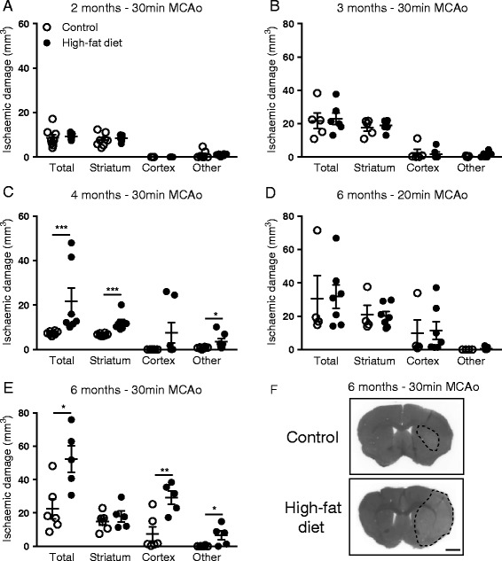
Male C57BL/6 mice were maintained on a high-fat (60% fat) or control (12% fat) diet for 2, 3, 4 and 6 months when experimental stroke was induced by transient occlusion of the middle cerebral artery (MCAo) for either 20 (6-month diet) or 30 min (2-, 3-, 4- and 6-month diet). Ischaemic damage, blood-brain barrier (BBB) integrity, neutrophil number and chemokine expression in the brain were assessed at 24 h. Plasma chemokine levels (at 4 and 24 h) and neutrophil number in the liver (at 24 h) were measured. Physiological parameters (body weight and blood glucose) were measured in naïve control- and high-fat-fed mice at all time points and blood pressure at 3 and 6 months. Blood cell counts were also assessed in naïve 6-month control- and high-fat-fed mice.
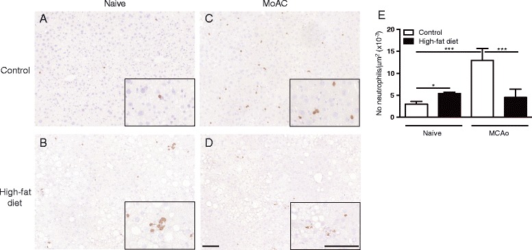
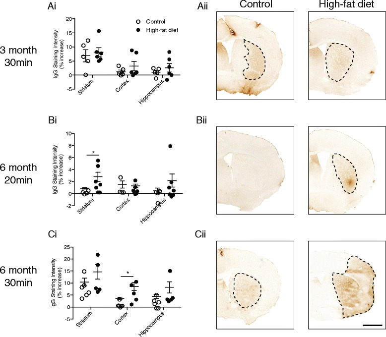
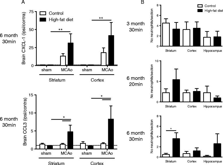
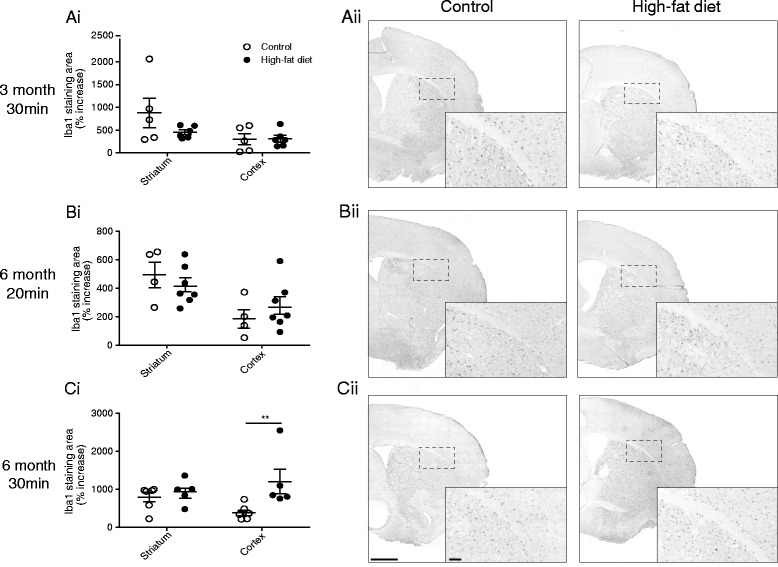
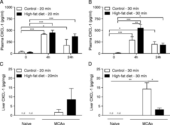
Mice fed a high-fat diet for 6 months had greater body weight, blood glucose and white and red blood cell count but no change in systolic blood pressure. After 4 and 6 months of high-fat feeding, and in the latter group with a 30-min (but not 20-min) occlusion of the MCA, obese mice had greater ischaemic brain damage. An increase in blood-brain barrier permeability, chemokine expression (CXCL-1 and CCL3), neutrophil number and microglia/macrophage cells was observed in the brains of 6-month high-fat-fed mice after 30-min MCAo. In response to stroke, chemokine (CXCL-1) expression in the plasma and liver was significantly different in obese mice (6-month high-fat fed), and a greater number of neutrophils were detected in the liver of control but not obese mice.
The detrimental effects of diet-induced obesity on stroke were therefore dependent on the severity of obesity and length of ischaemic challenge. The altered inflammatory response in obese mice may play a key role in its negative impact on stroke.
肥胖会增加缺血性中风的风险,并且在临床和实验中均与较差的预后相关。大多数实验研究使用的是肥胖的基因模型。在此,我们采用了一种更具临床相关性的模型——饮食诱导肥胖模型,来研究肥胖随时间推移对中风后预后和炎症反应的影响。
雄性C57BL/6小鼠分别给予高脂(60%脂肪)或对照(12%脂肪)饮食2、3、4和6个月,然后通过大脑中动脉短暂闭塞(MCAo)诱导实验性中风,闭塞时间在6个月饮食组为20分钟,在2、3、4和6个月饮食组为30分钟。在24小时时评估脑缺血损伤、血脑屏障(BBB)完整性、中性粒细胞数量和趋化因子表达。测量血浆趋化因子水平(4小时和24小时时)以及肝脏中的中性粒细胞数量(24小时时)。在所有时间点测量未处理的对照饮食和高脂饮食小鼠的生理参数(体重和血糖),并在3个月和6个月时测量血压。还评估了未处理的6个月对照饮食和高脂饮食小鼠的血细胞计数。
喂食高脂饮食6个月的小鼠体重、血糖以及白细胞和红细胞计数更高,但收缩压无变化。在高脂喂养4个月和6个月后,对于6个月饮食组中MCA闭塞30分钟(而非20分钟)的小鼠,肥胖小鼠的脑缺血损伤更大。在6个月高脂饮食小鼠经30分钟MCAo后,观察到其血脑屏障通透性增加、趋化因子表达(CXCL-1和CCL3)、中性粒细胞数量以及小胶质细胞/巨噬细胞数量增加。对中风的反应中,肥胖小鼠(6个月高脂饮食)血浆和肝脏中的趋化因子(CXCL-1)表达存在显著差异,并且在对照小鼠而非肥胖小鼠的肝脏中检测到更多的中性粒细胞。
因此,饮食诱导肥胖对中风的有害影响取决于肥胖的严重程度和缺血挑战的时长。肥胖小鼠中炎症反应的改变可能在其对中风的负面影响中起关键作用。