Rashid Arshad, Mushtaque Majid, Bali Rajandeep Singh, Nazir Saima, Khuroo Suhail, Ishaq Sheikh
Department of Health Services, Kashmir 190001, India.
Department of Surgery, Maulana Azad Medical College, New Delhi 110002, India.
Anat Res Int. 2015;2015:847812. doi: 10.1155/2015/847812. Epub 2015 Jul 9.
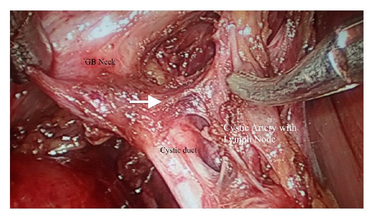
Uncontrolled arterial bleeding during laparoscopic cholecystectomy is a serious problem and may increase the risk of bile duct damage. Therefore, accurate identification of the anatomy of the cystic artery is very important. Cystic artery is notoriously known to have a highly variable branching pattern. We reviewed the anatomy of the cystic artery and its branch to cystic duct as seen through the video laparoscope. A single artery to cystic duct with the classical "H-configuration" was demonstrated in 161 (91.47%) patients. This branch may cause troublesome bleeding during laparoscopic dissection in the hepatobiliary triangle. Careful identification of artery to cystic duct is helpful in the proper dissection of Calot's triangle as it reduces the chances of hemorrhage and thus may also be helpful in prevention of extrahepatic biliary radical injuries.
腹腔镜胆囊切除术中未控制的动脉出血是一个严重问题,可能会增加胆管损伤的风险。因此,准确识别胆囊动脉的解剖结构非常重要。众所周知,胆囊动脉的分支模式高度可变。我们通过视频腹腔镜观察了胆囊动脉及其至胆囊管分支的解剖结构。161例(91.47%)患者显示有一条具有经典“H形结构”的至胆囊管的单一动脉。该分支在腹腔镜下解剖肝门三角时可能导致棘手的出血。仔细识别至胆囊管的动脉有助于正确解剖Calot三角,因为它减少了出血的机会,因此也可能有助于预防肝外胆管的根治性损伤。