• 文献检索
  • 文档翻译
  • 深度研究
  • 学术资讯
  • Suppr Zotero 插件Zotero 插件
  • 邀请有礼
  • 套餐&价格
  • 历史记录
应用&插件
Suppr Zotero 插件Zotero 插件浏览器插件Mac 客户端Windows 客户端微信小程序
定价
高级版会员购买积分包购买API积分包
服务
文献检索文档翻译深度研究API 文档MCP 服务
关于我们
关于 Suppr公司介绍联系我们用户协议隐私条款
关注我们

Suppr 超能文献

核心技术专利:CN118964589B侵权必究
粤ICP备2023148730 号-1Suppr @ 2026

文献检索

告别复杂PubMed语法,用中文像聊天一样搜索,搜遍4000万医学文献。AI智能推荐,让科研检索更轻松。

立即免费搜索

文件翻译

保留排版,准确专业,支持PDF/Word/PPT等文件格式,支持 12+语言互译。

免费翻译文档

深度研究

AI帮你快速写综述,25分钟生成高质量综述,智能提取关键信息,辅助科研写作。

立即免费体验

朊病毒蛋白N1和N2裂解片段与磷脂酰丝氨酸和磷脂酸结合;与应激保护反应的相关性。

The Prion Protein N1 and N2 Cleavage Fragments Bind to Phosphatidylserine and Phosphatidic Acid; Relevance to Stress-Protection Responses.

作者信息

Haigh Cathryn L, Tumpach Carolin, Drew Simon C, Collins Steven J

机构信息

Department of Medicine (Royal Melbourne Hospital), The University of Melbourne, Melbourne Brain Centre, Melbourne, VIC, AUS, 3010.

The Florey Department of Neuroscience and Mental Health, The University of Melbourne, Melbourne Brain Centre, Melbourne, VIC, AUS, 3010.

出版信息

PLoS One. 2015 Aug 7;10(8):e0134680. doi: 10.1371/journal.pone.0134680. eCollection 2015.

DOI:10.1371/journal.pone.0134680
PMID:26252007
原文链接:https://pmc.ncbi.nlm.nih.gov/articles/PMC4529310/
Abstract

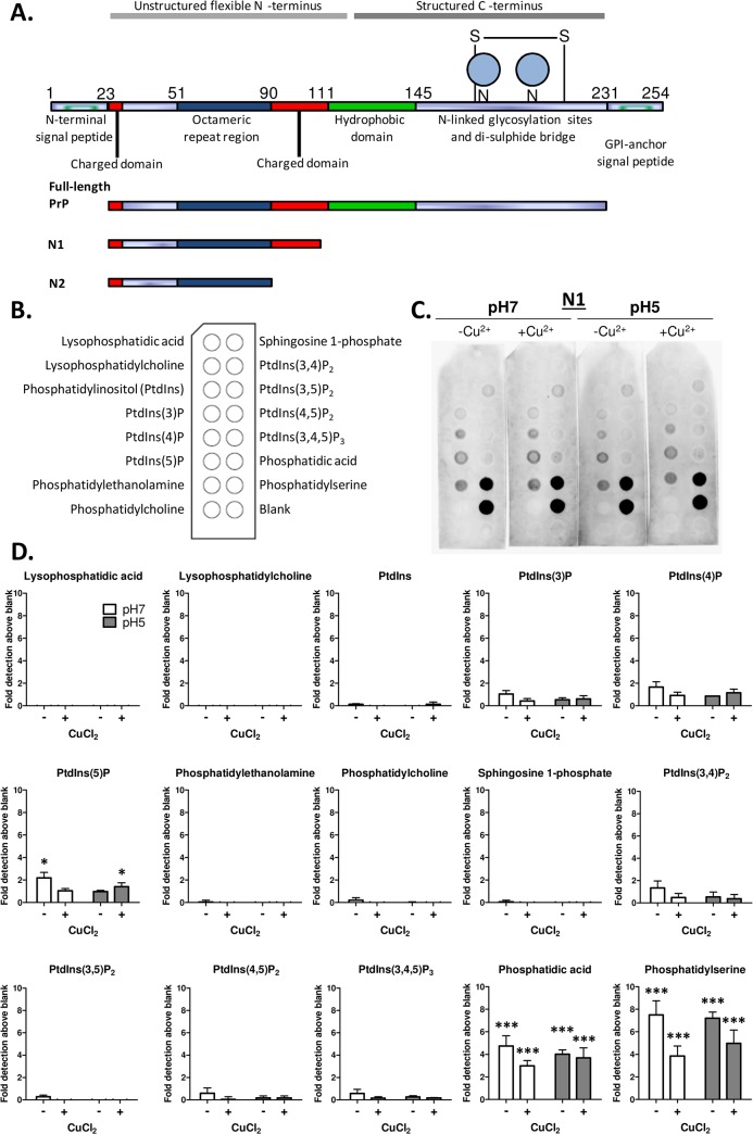
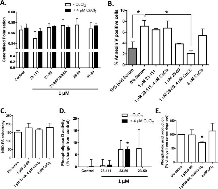
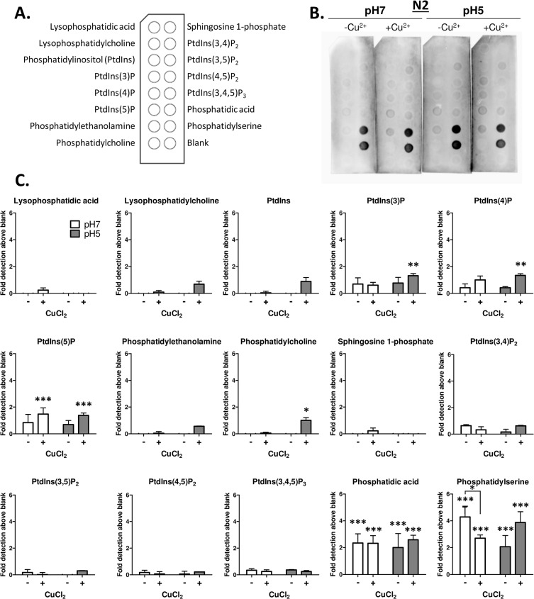
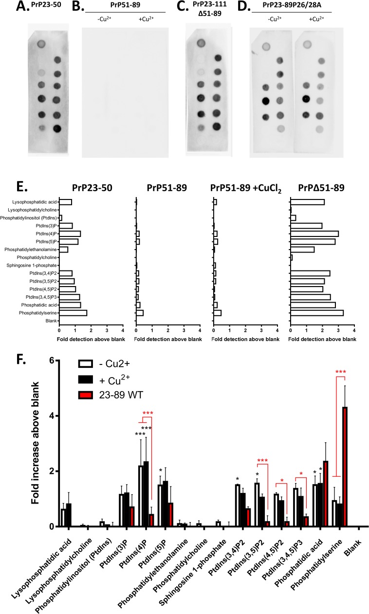
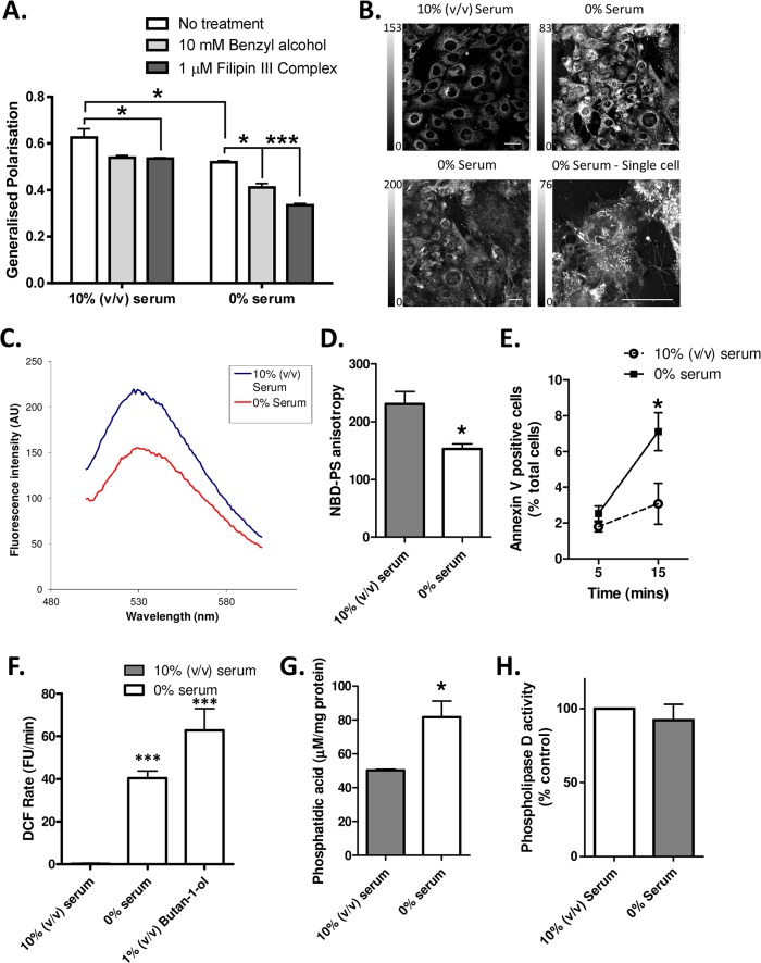
Internal cleavage of the cellular prion protein generates two well characterised N-terminal fragments, N1 and N2. These fragments have been shown to bind to anionic phospholipids at low pH. We sought to investigate binding with other lipid moieties and queried how such interactions could be relevant to the cellular functions of these fragments. Both N1 and N2 bound phosphatidylserine (PS), as previously reported, and a further interaction with phosphatidic acid (PA) was also identified. The specificity of this interaction required the N-terminus, especially the proline motif within the basic amino acids at the N-terminus, together with the copper-binding region (unrelated to copper saturation). Previously, the fragments have been shown to be protective against cellular stresses. In the current study, serum deprivation was used to induce changes in the cellular lipid environment, including externalisation of plasma membrane PS and increased cellular levels of PA. When copper-saturated, N2 could reverse these changes, but N1 could not, suggesting that direct binding of N2 to cellular lipids may be part of the mechanism by which this peptide signals its protective response.

摘要

细胞朊蛋白的内源性切割产生了两个特征明确的N端片段,即N1和N2。这些片段已被证明在低pH值下能与阴离子磷脂结合。我们试图研究它们与其他脂质部分的结合情况,并探讨这种相互作用与这些片段的细胞功能有何关联。如先前报道,N1和N2都能结合磷脂酰丝氨酸(PS),并且还发现了它们与磷脂酸(PA)的进一步相互作用。这种相互作用的特异性需要N端,特别是N端碱性氨基酸内的脯氨酸基序,以及铜结合区域(与铜饱和度无关)。此前,这些片段已被证明对细胞应激具有保护作用。在当前研究中,血清剥夺被用于诱导细胞脂质环境的变化,包括质膜PS的外化和细胞内PA水平的升高。当铜饱和时,N2可以逆转这些变化,但N1不能,这表明N2与细胞脂质的直接结合可能是该肽发出其保护反应信号的机制的一部分。

https://cdn.ncbi.nlm.nih.gov/pmc/blobs/438f/4529310/cf8615e20118/pone.0134680.g005.jpg
https://cdn.ncbi.nlm.nih.gov/pmc/blobs/438f/4529310/5130503f95a9/pone.0134680.g001.jpg
https://cdn.ncbi.nlm.nih.gov/pmc/blobs/438f/4529310/5ca1449b0625/pone.0134680.g002.jpg
https://cdn.ncbi.nlm.nih.gov/pmc/blobs/438f/4529310/3cc3e9e669b7/pone.0134680.g003.jpg
https://cdn.ncbi.nlm.nih.gov/pmc/blobs/438f/4529310/d845a96869a7/pone.0134680.g004.jpg
https://cdn.ncbi.nlm.nih.gov/pmc/blobs/438f/4529310/cf8615e20118/pone.0134680.g005.jpg

相似文献

1
The Prion Protein N1 and N2 Cleavage Fragments Bind to Phosphatidylserine and Phosphatidic Acid; Relevance to Stress-Protection Responses.朊病毒蛋白N1和N2裂解片段与磷脂酰丝氨酸和磷脂酸结合;与应激保护反应的相关性。
PLoS One. 2015 Aug 7;10(8):e0134680. doi: 10.1371/journal.pone.0134680. eCollection 2015.
2
Neutron reflectometry studies define prion protein N-terminal peptide membrane binding.中子反射率研究确定了朊病毒蛋白N端肽的膜结合情况。
Biophys J. 2014 Nov 18;107(10):2313-24. doi: 10.1016/j.bpj.2014.09.027.
3
The catalytic domains of Clostridium sordellii lethal toxin and related large clostridial glucosylating toxins specifically recognize the negatively charged phospholipids phosphatidylserine and phosphatidic acid.索氏梭菌致死毒素及相关的大型梭菌糖基化毒素的催化结构域可特异性识别带负电荷的磷脂——磷脂酰丝氨酸和磷脂酸。
Cell Microbiol. 2015 Oct;17(10):1477-93. doi: 10.1111/cmi.12449. Epub 2015 May 13.
4
Dominant roles of the polybasic proline motif and copper in the PrP23-89-mediated stress protection response.多碱性脯氨酸基序和铜在PrP23-89介导的应激保护反应中的主导作用。
J Cell Sci. 2009 May 15;122(Pt 10):1518-28. doi: 10.1242/jcs.043604. Epub 2009 Apr 21.
5
Interaction of the Spo20 membrane-sensor motif with phosphatidic acid and other anionic lipids, and influence of the membrane environment.Spo20膜传感器基序与磷脂酸及其他阴离子脂质的相互作用以及膜环境的影响。
PLoS One. 2014 Nov 26;9(11):e113484. doi: 10.1371/journal.pone.0113484. eCollection 2014.
6
Membrane interactions and conformational preferences of human and avian prion N-terminal tandem repeats: the role of copper(II) ions, pH, and membrane mimicking environments.人源和禽源朊病毒 N 端串联重复序列的膜相互作用和构象偏好:铜(II)离子、pH 值和膜模拟环境的作用。
J Phys Chem B. 2010 Nov 4;114(43):13830-8. doi: 10.1021/jp1033036.
7
Anionic phospholipid interactions of the prion protein N terminus are minimally perturbing and not driven solely by the octapeptide repeat domain.朊病毒蛋白 N 端的阴离子磷脂相互作用的干扰极小,并非仅由八肽重复结构域驱动。
J Biol Chem. 2010 Oct 15;285(42):32282-92. doi: 10.1074/jbc.M110.123398. Epub 2010 Aug 2.
8
PrPC-related signal transduction is influenced by copper, membrane integrity and the alpha cleavage site.与朊蛋白(PrPC)相关的信号转导受铜、膜完整性和α裂解位点的影响。
Cell Res. 2009 Sep;19(9):1062-78. doi: 10.1038/cr.2009.86. Epub 2009 Jul 14.
9
The carboxy-terminal tail domain of vinculin contains a cryptic binding site for acidic phospholipids.纽蛋白的羧基末端尾域含有一个对酸性磷脂的隐蔽结合位点。
Biochem Biophys Res Commun. 1995 May 5;210(1):159-64. doi: 10.1006/bbrc.1995.1641.
10
Phosphatidic acid plays key roles regulating plant development and stress responses.磷脂酸在调节植物发育和应激反应方面发挥着关键作用。
J Integr Plant Biol. 2018 Sep;60(9):851-863. doi: 10.1111/jipb.12655. Epub 2018 Jul 9.

引用本文的文献

1
Prion Protein Endoproteolysis: Cleavage Sites, Mechanisms and Connections to Prion Disease.朊病毒蛋白内蛋白水解作用:切割位点、机制及其与朊病毒疾病的关联
J Neurochem. 2025 Jan;169(1):e16310. doi: 10.1111/jnc.16310.
2
What is the role of lipids in prion conversion and disease?脂质在朊病毒转化和疾病中起什么作用?
Front Mol Neurosci. 2023 Jan 10;15:1032541. doi: 10.3389/fnmol.2022.1032541. eCollection 2022.
3
Minimal change prion retinopathy: Morphometric comparison of retinal and brain prion deposits in Creutzfeldt-Jakob disease.

本文引用的文献

1
Neutron reflectometry studies define prion protein N-terminal peptide membrane binding.中子反射率研究确定了朊病毒蛋白N端肽的膜结合情况。
Biophys J. 2014 Nov 18;107(10):2313-24. doi: 10.1016/j.bpj.2014.09.027.
2
MEK1 transduces the prion protein N2 fragment antioxidant effects.丝裂原活化蛋白激酶激酶 1(MEK1)转导朊病毒蛋白 N2 片段的抗氧化作用。
Cell Mol Life Sci. 2015 Apr;72(8):1613-29. doi: 10.1007/s00018-014-1777-y. Epub 2014 Nov 13.
3
The role of diacylglycerol kinase ζ and phosphatidic acid in the mechanical activation of mammalian target of rapamycin (mTOR) signaling and skeletal muscle hypertrophy.
微小病变朊病毒视网膜病变:克雅氏病中视网膜和脑部朊病毒沉积的形态计量学比较。
Exp Eye Res. 2022 Sep;222:109172. doi: 10.1016/j.exer.2022.109172. Epub 2022 Jul 6.
4
Prion Protein: The Molecule of Many Forms and Faces.朊病毒蛋白:形态多样的分子。
Int J Mol Sci. 2022 Jan 22;23(3):1232. doi: 10.3390/ijms23031232.
5
β-Cleavage of the prion protein in the human eye: Implications for the spread of infectious prions and human ocular disorders.人眼中朊病毒蛋白的β 裂解:对传染性朊病毒传播和人类眼部疾病的影响。
Exp Eye Res. 2021 Nov;212:108787. doi: 10.1016/j.exer.2021.108787. Epub 2021 Oct 7.
6
Harnessing the Physiological Functions of Cellular Prion Protein in the Kidneys: Applications for Treating Renal Diseases.利用肾脏中细胞朊病毒蛋白的生理功能:治疗肾脏疾病的应用。
Biomolecules. 2021 May 22;11(6):784. doi: 10.3390/biom11060784.
7
N-Terminal Regions of Prion Protein: Functions and Roles in Prion Diseases.朊病毒蛋白的 N 端结构域:在朊病毒病中的功能和作用。
Int J Mol Sci. 2020 Aug 28;21(17):6233. doi: 10.3390/ijms21176233.
8
Show Me Your Friends and I Tell You Who You Are: The Many Facets of Prion Protein in Stroke.物以类聚,人以群分:朊病毒蛋白在卒中中的多面性。
Cells. 2020 Jul 2;9(7):1609. doi: 10.3390/cells9071609.
9
Prion protein N1 cleavage peptides stimulate microglial interaction with surrounding cells.朊病毒蛋白 N1 裂解肽刺激小胶质细胞与周围细胞的相互作用。
Sci Rep. 2020 Apr 20;10(1):6654. doi: 10.1038/s41598-020-63472-z.
10
Prion protein cleavage fragments regulate adult neural stem cell quiescence through redox modulation of mitochondrial fission and SOD2 expression.朊病毒蛋白裂解片段通过氧化还原调节线粒体分裂和 SOD2 表达来调控成年神经干细胞静止。
Cell Mol Life Sci. 2018 Sep;75(17):3231-3249. doi: 10.1007/s00018-018-2790-3. Epub 2018 Mar 24.
二酰基甘油激酶 ζ 和磷脂酸在机械激活哺乳动物雷帕霉素靶蛋白 (mTOR) 信号和骨骼肌肥大中的作用。
J Biol Chem. 2014 Jan 17;289(3):1551-63. doi: 10.1074/jbc.M113.531392. Epub 2013 Dec 3.
4
Cofactor molecules induce structural transformation during infectious prion formation.辅助因子分子在传染性朊病毒形成过程中诱导结构转化。
Structure. 2013 Nov 5;21(11):2061-8. doi: 10.1016/j.str.2013.08.025. Epub 2013 Oct 10.
5
Cytosolic caspases mediate mislocalised SOD2 depletion in an in vitro model of chronic prion infection.细胞溶质胱天蛋白酶介导慢性朊病毒感染体外模型中 SOD2 的定位缺失。
Dis Model Mech. 2013 Jul;6(4):952-63. doi: 10.1242/dmm.010678. Epub 2013 Apr 4.
6
PrP(C) homodimerization stimulates the production of PrPC cleaved fragments PrPN1 and PrPC1.朊病毒蛋白 C(PrP(C))同源二聚体刺激产生朊病毒蛋白 C 切割片段 PrPN1 和 PrPC1。
J Neurosci. 2012 Sep 19;32(38):13255-63. doi: 10.1523/JNEUROSCI.2236-12.2012.
7
Lipid rafts generate digital-like signal transduction in cell plasma membranes.脂筏在细胞质膜中产生数字样信号转导。
Biotechnol J. 2012 Jun;7(6):753-61. doi: 10.1002/biot.201100360. Epub 2012 Apr 5.
8
The extracellular regulated kinase-1 (ERK1) controls regulated alpha-secretase-mediated processing, promoter transactivation, and mRNA levels of the cellular prion protein.细胞外调节激酶 1(ERK1)控制调节的 α-分泌酶介导的加工、启动子转录激活和朊病毒蛋白的 mRNA 水平。
J Biol Chem. 2011 Aug 19;286(33):29192-29206. doi: 10.1074/jbc.M110.208249. Epub 2011 May 17.
9
Acute exposure to prion infection induces transient oxidative stress progressing to be cumulatively deleterious with chronic propagation in vitro.急性朊病毒感染会引发短暂的氧化应激,这种应激在体外的慢性传播过程中会逐渐累积,造成损害。
Free Radic Biol Med. 2011 Aug 1;51(3):594-608. doi: 10.1016/j.freeradbiomed.2011.03.035. Epub 2011 Apr 3.
10
Optical imaging detects apoptosis in the brain and peripheral organs of prion-infected mice.光学成像是一种可以在感染朊病毒的老鼠的大脑和外周器官中检测细胞凋亡的方法。
J Neuropathol Exp Neurol. 2011 Feb;70(2):143-50. doi: 10.1097/NEN.0b013e3182084a8c.