• 文献检索
  • 文档翻译
  • 深度研究
  • 学术资讯
  • Suppr Zotero 插件Zotero 插件
  • 邀请有礼
  • 套餐&价格
  • 历史记录
应用&插件
Suppr Zotero 插件Zotero 插件浏览器插件Mac 客户端Windows 客户端微信小程序
定价
高级版会员购买积分包购买API积分包
服务
文献检索文档翻译深度研究API 文档MCP 服务
关于我们
关于 Suppr公司介绍联系我们用户协议隐私条款
关注我们

Suppr 超能文献

核心技术专利:CN118964589B侵权必究
粤ICP备2023148730 号-1Suppr @ 2026

文献检索

告别复杂PubMed语法,用中文像聊天一样搜索,搜遍4000万医学文献。AI智能推荐,让科研检索更轻松。

立即免费搜索

文件翻译

保留排版,准确专业,支持PDF/Word/PPT等文件格式,支持 12+语言互译。

免费翻译文档

深度研究

AI帮你快速写综述,25分钟生成高质量综述,智能提取关键信息,辅助科研写作。

立即免费体验

使用3D MRI对乳腺四个象限的局部乳腺密度进行量化——一项初步研究

Quantification of Regional Breast Density in Four Quadrants Using 3D MRI-A Pilot Study.

作者信息

Fwu Peter T, Chen Jeon-Hor, Li Yifan, Chan Siwa, Su Min-Ying

机构信息

Center for Functional Onco-Imaging, Department of Radiological Sciences, University of California, Irvine, CA, USA.

Center for Functional Onco-Imaging, Department of Radiological Sciences, University of California, Irvine, CA, USA; Department of Radiology, E-Da Hospital and I-Shou University, Kaohsiung, Taiwan.

出版信息

Transl Oncol. 2015 Aug;8(4):250-7. doi: 10.1016/j.tranon.2015.04.005.

DOI:10.1016/j.tranon.2015.04.005
PMID:26310370
原文链接:https://pmc.ncbi.nlm.nih.gov/articles/PMC4562976/
Abstract

PURPOSE

This study presented a three-dimensional magnetic resonance (MR)-based method to separate a breast into four quadrants for quantitative measurements of the quadrant breast volume (BV) and density.

METHODS

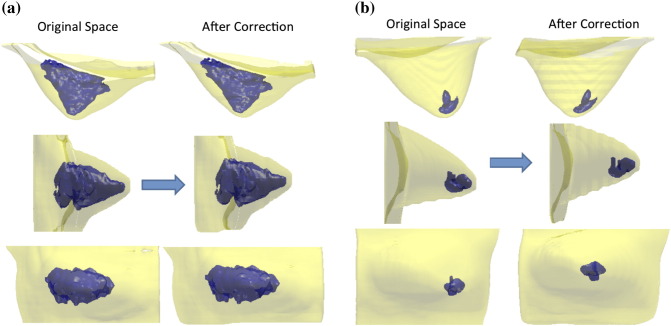
Breast MR images from 58 healthy women were studied. The breast and the fibroglandular tissue were segmented by using a computer-based algorithm. A breast was divided into four quadrants using two perpendicular planes intersecting at the nipple or the nipple-centroid line. After the separation, the BV, the fibroglandular tissue volume, and the percent density (PD) were calculated. The symmetry of the quadrant BV in the left and right breasts separated by using the nipple alone, or the nipple-centroid line, was compared.

RESULTS

The quadrant separation made on the basis of the nipple-centroid line showed closer BVs in four quadrants than using the nipple alone. The correlation and agreement for the BV in corresponding quadrants of the left and the right breasts were improved after the nipple-centroid reorientation. Among the four quadrants, PD was the highest in the lower outer and the lowest in the upper outer (significant than the other three) quadrants (P < .05).

CONCLUSIONS

We presented a quantitative method to divide a breast into four quadrants. The reorientation based on the nipple-centroid line improved the left to right quadrant symmetry, and this may provide a better standardized method to measure quantitative quadrant density. The cancer occurrence rates are known to vary in different sites of a breast, and our method may provide a tool for investigating its association with the quantitative breast density.

摘要

目的

本研究提出了一种基于三维磁共振(MR)的方法,将乳房分为四个象限,以定量测量象限乳房体积(BV)和密度。

方法

对58名健康女性的乳房MR图像进行研究。使用基于计算机的算法对乳房和纤维腺体组织进行分割。通过两个在乳头或乳头-质心线处相交的垂直平面将乳房分为四个象限。分割后,计算BV、纤维腺体组织体积和百分比密度(PD)。比较仅使用乳头或乳头-质心线分割的左右乳房象限BV的对称性。

结果

基于乳头-质心线进行的象限分割显示,四个象限中的BV比仅使用乳头时更接近。乳头-质心重新定位后,左右乳房相应象限中BV的相关性和一致性得到改善。在四个象限中,PD在乳房外下象限最高,在上外象限最低(显著低于其他三个象限)(P <.05)。

结论

我们提出了一种将乳房分为四个象限的定量方法。基于乳头-质心线的重新定位改善了左右象限的对称性,这可能为测量定量象限密度提供更好的标准化方法。已知乳腺癌发生率在乳房的不同部位有所不同,我们的方法可能为研究其与定量乳房密度的关联提供一种工具。

https://cdn.ncbi.nlm.nih.gov/pmc/blobs/7d6b/4562976/06b85d952b69/gr4.jpg
https://cdn.ncbi.nlm.nih.gov/pmc/blobs/7d6b/4562976/10a566af3a98/gr1.jpg
https://cdn.ncbi.nlm.nih.gov/pmc/blobs/7d6b/4562976/88df2edab69c/gr2.jpg
https://cdn.ncbi.nlm.nih.gov/pmc/blobs/7d6b/4562976/3a4e99830ca8/gr3.jpg
https://cdn.ncbi.nlm.nih.gov/pmc/blobs/7d6b/4562976/06b85d952b69/gr4.jpg
https://cdn.ncbi.nlm.nih.gov/pmc/blobs/7d6b/4562976/10a566af3a98/gr1.jpg
https://cdn.ncbi.nlm.nih.gov/pmc/blobs/7d6b/4562976/88df2edab69c/gr2.jpg
https://cdn.ncbi.nlm.nih.gov/pmc/blobs/7d6b/4562976/3a4e99830ca8/gr3.jpg
https://cdn.ncbi.nlm.nih.gov/pmc/blobs/7d6b/4562976/06b85d952b69/gr4.jpg

相似文献

1
Quantification of Regional Breast Density in Four Quadrants Using 3D MRI-A Pilot Study.使用3D MRI对乳腺四个象限的局部乳腺密度进行量化——一项初步研究
Transl Oncol. 2015 Aug;8(4):250-7. doi: 10.1016/j.tranon.2015.04.005.
2
3D MRI for Quantitative Analysis of Quadrant Percent Breast Density: Correlation with Quadrant Location of Breast Cancer.用于乳腺象限百分比密度定量分析的3D磁共振成像:与乳腺癌象限位置的相关性
Acad Radiol. 2017 Jul;24(7):811-817. doi: 10.1016/j.acra.2016.12.016. Epub 2017 Jan 26.
3
Evaluation of the association between quantitative mammographic density and breast cancer occurred in different quadrants.评估不同象限乳腺钼靶定量密度与乳腺癌之间的关联。
BMC Cancer. 2017 Apr 17;17(1):274. doi: 10.1186/s12885-017-3270-0.
4
Impact of positional difference on the measurement of breast density using MRI.体位差异对使用MRI测量乳腺密度的影响。
Med Phys. 2015 May;42(5):2268-75. doi: 10.1118/1.4917083.
5
Response of bilateral breasts to the endogenous hormonal fluctuation in a menstrual cycle evaluated using 3D MRI.经 3D MRI 评估,双侧乳房对月经周期内内源性激素波动的反应。
Magn Reson Imaging. 2013 May;31(4):538-44. doi: 10.1016/j.mri.2012.10.022. Epub 2012 Dec 5.
6
Opportunistic Breast Density Assessment in Women Receiving Low-dose Chest Computed Tomography Screening.接受低剂量胸部计算机断层扫描筛查的女性的机会性乳腺密度评估
Acad Radiol. 2016 Sep;23(9):1154-61. doi: 10.1016/j.acra.2016.05.003. Epub 2016 Jun 6.
7
Fully automated nipple detection in digital breast tomosynthesis.数字乳腺断层合成中的全自动乳头检测
Comput Methods Programs Biomed. 2017 May;143:113-120. doi: 10.1016/j.cmpb.2017.03.004. Epub 2017 Mar 6.
8
Correlation between mammographic density and volumetric fibroglandular tissue estimated on breast MR images.乳腺钼靶密度与乳腺磁共振成像估计的纤维腺体组织体积之间的相关性。
Med Phys. 2004 Apr;31(4):933-42. doi: 10.1118/1.1668512.
9
Circumareolar Incision-subdermal Tunneling Dissection for Excision of Multiple Breast Fibroadenomata.乳晕周围切口-皮下隧道剥离术用于多发性乳腺纤维瘤切除
Niger J Surg. 2017 Jan-Jun;23(1):63-66. doi: 10.4103/1117-6806.205749.
10
Sections of the nipple and quadrants in mastectomy specimens for carcinoma are of limited value.乳腺癌乳房切除标本中的乳头及象限切片价值有限。
J Clin Pathol. 2005 May;58(5):543-5. doi: 10.1136/jcp.2004.022665.

引用本文的文献

1
Quantitative Study on the Breast Density and the Volume of the Mammary Gland According to the Patient's Age and Breast Quadrant.根据患者年龄和乳房象限对乳房密度及乳腺体积的定量研究
Diagnostics (Basel). 2023 Oct 30;13(21):3343. doi: 10.3390/diagnostics13213343.
2
Automatic and fast segmentation of breast region-of-interest (ROI) and density in MRIs.磁共振成像中乳腺感兴趣区域(ROI)及密度的自动快速分割
Heliyon. 2018 Dec 17;4(12):e01042. doi: 10.1016/j.heliyon.2018.e01042. eCollection 2018 Dec.
3
Evaluation of the association between quantitative mammographic density and breast cancer occurred in different quadrants.

本文引用的文献

1
Breast tissue characterization with photon-counting spectral CT imaging: a postmortem breast study.基于光子计数光谱CT成像的乳腺组织特征分析:一项乳腺尸体研究
Radiology. 2014 Sep;272(3):731-8. doi: 10.1148/radiol.14132732. Epub 2014 May 7.
2
Template-based automatic breast segmentation on MRI by excluding the chest region.基于模板的自动 MRI 乳房分割,排除胸部区域。
Med Phys. 2013 Dec;40(12):122301. doi: 10.1118/1.4828837.
3
Consistency of breast density measured from the same women in four different MR scanners.在四台不同的磁共振成像扫描仪上对同一女性测量的乳腺密度的一致性。
评估不同象限乳腺钼靶定量密度与乳腺癌之间的关联。
BMC Cancer. 2017 Apr 17;17(1):274. doi: 10.1186/s12885-017-3270-0.
4
3D MRI for Quantitative Analysis of Quadrant Percent Breast Density: Correlation with Quadrant Location of Breast Cancer.用于乳腺象限百分比密度定量分析的3D磁共振成像:与乳腺癌象限位置的相关性
Acad Radiol. 2017 Jul;24(7):811-817. doi: 10.1016/j.acra.2016.12.016. Epub 2017 Jan 26.
Med Phys. 2012 Aug;39(8):4886-95. doi: 10.1118/1.4736824.
4
Comparison of breast density measured on MR images acquired using fat-suppressed versus nonfat-suppressed sequences.比较使用脂肪抑制和非脂肪抑制序列获取的 MR 图像上测量的乳腺密度。
Med Phys. 2011 Nov;38(11):5961-8. doi: 10.1118/1.3646756.
5
Menstrual cycle-related fluctuations in breast density measured by using three-dimensional MR imaging.采用三维磁共振成像测量的与月经周期相关的乳腺密度变化。
Radiology. 2011 Dec;261(3):744-51. doi: 10.1148/radiol.11110506. Epub 2011 Aug 30.
6
Tissue composition of mammographically dense and non-dense breast tissue.乳腺组织中致密型和非致密型组织的构成。
Breast Cancer Res Treat. 2012 Jan;131(1):267-75. doi: 10.1007/s10549-011-1727-4. Epub 2011 Aug 30.
7
Objective breast symmetry evaluation using 3-D surface imaging.三维表面成像在乳房对称性评估中的应用。
Breast. 2012 Apr;21(2):152-8. doi: 10.1016/j.breast.2011.07.016. Epub 2011 Aug 17.
8
Localized fibroglandular tissue as a predictor of future tumor location within the breast.局部纤维腺体组织可预测乳腺内未来肿瘤的位置。
Cancer Epidemiol Biomarkers Prev. 2011 Aug;20(8):1718-25. doi: 10.1158/1055-9965.EPI-11-0423. Epub 2011 Jun 21.
9
Computerized prediction of risk for developing breast cancer based on bilateral mammographic breast tissue asymmetry.基于双侧乳腺组织不对称的计算机预测乳腺癌发病风险。
Med Eng Phys. 2011 Oct;33(8):934-42. doi: 10.1016/j.medengphy.2011.03.001. Epub 2011 Apr 8.
10
Lymphatic drainage and tumour prevalence in the breast: a statistical analysis of symmetry, gender and node field independence.淋巴引流与乳房肿瘤患病率:对称性、性别与淋巴结场独立性的统计学分析。
J Anat. 2011 Jun;218(6):652-9. doi: 10.1111/j.1469-7580.2011.01362.x. Epub 2011 Mar 31.