Glimm A-M, Werner S G, Burmester G R, Backhaus M, Ohrndorf S
Department of Rheumatology and Clinical Immunology, Charité Universitätsmedizin Berlin, Berlin, Germany.
Ann Rheum Dis. 2016 Mar;75(3):566-70. doi: 10.1136/annrheumdis-2015-207345. Epub 2015 Aug 26.
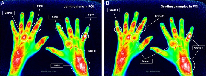
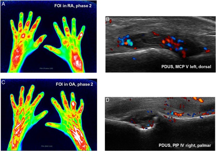
In rheumatoid arthritis (RA), hand synovitis appears especially in wrist, metacarpophalangeal (MCP) and proximal interphalangeal (PIP) joints. In hand osteoarthritis (OA), potential inflammatory changes are mainly present in PIP and distal interphalangeal (DIP) joints. Joint inflammation can be visualised by fluorescence optical imaging (FOI) and musculoskeletal ultrasound (US).
Comparison of the amount and distribution of inflammatory signs in wrist and finger joints of the clinically dominant hand in patients with OA and RA by FOI and gray-scale (GSUS) and power Doppler US (PDUS).
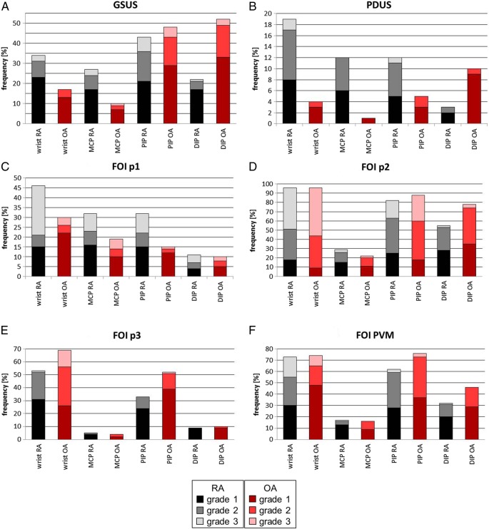
FOI and GSUS/PDUS were performed in 1.170 joints (wrists, MCP, PIP, DIP) in 90 patients (67 RA, 23 OA). Joint inflammation was graded by a semiquantitative score (0-3) for each imaging method.
GSUS/PDUS showed wrist and MCP joints mostly affected in RA. DIP joints were graded higher in OA. In FOI, RA and OA featured inflammatory changes in the respective joint groups depending on the phase of fluorescence dye flooding.
US and FOI detected inflammation in both RA and OA highlighting the inflammatory component in the course of OA. The different inflammatory patterns and various shapes of fluorescence enhancement in FOI may offer opportunities to distinguish and determine the inflammatory status in both diseases.
在类风湿关节炎(RA)中,手部滑膜炎尤其出现在腕关节、掌指关节(MCP)和近端指间关节(PIP)。在手部骨关节炎(OA)中,潜在的炎症变化主要存在于PIP和远端指间关节(DIP)。关节炎症可通过荧光光学成像(FOI)和肌肉骨骼超声(US)进行可视化。
通过FOI、灰阶超声(GSUS)和能量多普勒超声(PDUS)比较OA和RA患者临床优势手的腕关节和手指关节炎症体征的数量和分布。
对90例患者(67例RA,23例OA)的1170个关节(腕关节、MCP、PIP、DIP)进行FOI和GSUS/PDUS检查。每种成像方法通过半定量评分(0-3)对关节炎症进行分级。
GSUS/PDUS显示RA中腕关节和MCP关节受累最严重。OA中DIP关节分级更高。在FOI中,RA和OA在各自的关节组中根据荧光染料充盈阶段呈现炎症变化。
US和FOI在RA和OA中均检测到炎症,突出了OA病程中的炎症成分。FOI中不同的炎症模式和荧光增强的各种形态可能为区分和确定两种疾病中的炎症状态提供机会。