Rheumatology, Immunology, Osteology Center, Duesseldorf, Germany.
Ann Rheum Dis. 2012 Apr;71(4):504-10. doi: 10.1136/annrheumdis-2010-148288.
Indocyanine green (ICG)-enhanced fluorescence optical imaging (FOI) is an established technology for imaging of inflammation in animal models. In experimental models of arthritis, FOI findings corresponded to histologically proven synovitis. This is the first comparative study of FOI with other imaging modalities in humans with arthritis.
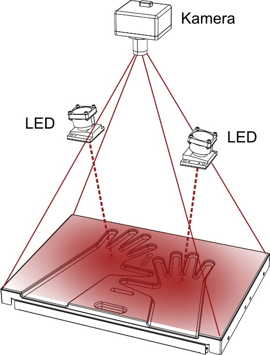
252 FOI examinations (Xiralite system, mivenion GmbH, Berlin, Germany; ICG bolus of 0.1 mg/kg/body weight, sequence of 360 images, one image per second) were compared with clinical examination (CE), ultrasonography (US) and MRI of patients with arthritis of the hands.
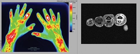
In an FOI sequence, three phases could be distinguished (P1-P3). With MRI as reference, FOI had a sensitivity of 76% and a specificity of 54%, while the specificity of phase 1 was 94%. FOI had agreement rates up to 88% versus CE, 64% versus greyscale US, 88% versus power Doppler US and 83% versus MRI, depending on the compared phase and parameter. FOI showed a higher rate of positive results compared to CE, US and MRI. In individual patients, FOI correlated significantly (p<0.05) with disease activity (Disease Activity Score 28, r=0.41), US (r=0.40) and RAMRIS (Rheumatoid Arthritis MRI Score) (r=0.56). FOI was normal in 97.8% of joints of controls.
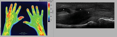
ICG-enhanced FOI is a new technology offering sensitive imaging detection of inflammatory changes in subjects with arthritis. FOI was more sensitive than CE and had good agreement with CE, US in power Doppler mode and MRI, while showing more positive results than these. An adequate interpretation of an FOI sequence requires a separate evaluation of all phases. For the detection of synovitis and tenosynovitis, FOI appears to be as informative as 1.5 T MRI and US.
吲哚菁绿(ICG)增强荧光光学成像(FOI)是一种用于动物模型炎症成像的成熟技术。在关节炎的实验模型中,FOI 结果与组织学证实的滑膜炎相对应。这是首次在人类关节炎患者中对 FOI 与其他成像方式进行比较的研究。
对 252 例 FOI 检查(Xiralite 系统,mivenion GmbH,柏林,德国;ICG 推注量为 0.1mg/kg/体重,360 张图像序列,每秒一张图像)与关节炎患者的临床检查(CE)、超声(US)和 MRI 进行比较。
在 FOI 序列中,可以区分三个阶段(P1-P3)。以 MRI 为参考,FOI 的灵敏度为 76%,特异性为 54%,而第 1 阶段的特异性为 94%。FOI 与 CE 的符合率高达 88%,与灰阶 US 的符合率为 64%,与功率多普勒 US 的符合率为 88%,与 MRI 的符合率为 83%,具体取决于比较的阶段和参数。与 CE、US 和 MRI 相比,FOI 显示出更高的阳性结果率。在个别患者中,FOI 与疾病活动度(28 关节疾病活动度评分,r=0.41)、US(r=0.40)和 RAMRIS(类风湿关节炎 MRI 评分,r=0.56)显著相关(p<0.05)。对照组的 97.8%关节 FOI 正常。
ICG 增强 FOI 是一种新的技术,可敏感地检测关节炎患者的炎症变化。FOI 比 CE 更敏感,与 CE、功率多普勒模式下的 US 和 MRI 的一致性较好,而阳性结果比这些检查更多。要对 FOI 序列进行充分的解读,需要单独评估所有阶段。对于滑膜炎和腱鞘炎的检测,FOI 似乎与 1.5T MRI 和 US 同样具有信息量。