Stoddart Leigh A, White Carl W, Nguyen Kim, Hill Stephen J, Pfleger Kevin D G
Cell Signalling Research Group, School of Life Sciences, University of Nottingham, Nottingham, UK.
Molecular Endocrinology and Pharmacology, Harry Perkins Institute of Medical Research, Nedlands, WA, Australia.
Br J Pharmacol. 2016 Oct;173(20):3028-37. doi: 10.1111/bph.13316. Epub 2015 Nov 5.
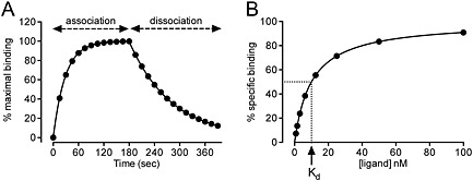
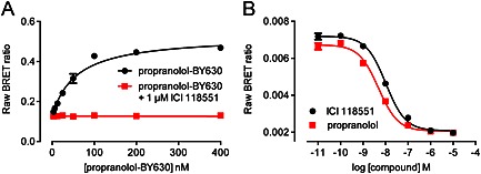
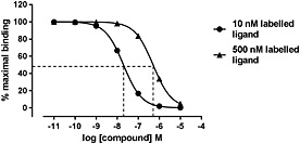
Ligand binding is a vital component of any pharmacologist's toolbox and allows the detailed investigation of how a molecule binds to its receptor. These studies enable the experimental determination of binding affinity of labelled and unlabelled compounds through kinetic, saturation (Kd ) and competition (Ki ) binding assays. Traditionally, these studies have used molecules labelled with radioisotopes; however, more recently, fluorescent ligands have been developed for this purpose. This review will briefly cover receptor ligand binding theory and then discuss the use of fluorescent ligands with some of the different technologies currently employed to examine ligand binding. Fluorescent ligands can be used for direct measurement of receptor-associated fluorescence using confocal microscopy and flow cytometry as well as in assays such as fluorescence polarization, where ligand binding is monitored by changes in the free rotation when a fluorescent ligand is bound to a receptor. Additionally, fluorescent ligands can act as donors or acceptors for fluorescence resonance energy transfer (FRET) with the development of assays based on FRET and time-resolved FRET (TR-FRET). Finally, we have recently developed a novel bioluminescence resonance energy transfer (BRET) ligand binding assay utilizing a small (19 kDa), super-bright luciferase subunit (NanoLuc) from a deep sea shrimp. In combination with fluorescent ligands, measurement of RET now provides an array of methodologies to study ligand binding. While each method has its own advantages and drawbacks, binding studies using fluorescent ligands are now a viable alternative to the use of radioligands. Linked Articles This article is part of a themed section on Molecular Pharmacology of G Protein-Coupled Receptors. To view the other articles in this section visit http://onlinelibrary.wiley.com/doi/10.1111/bph.v173.20/issuetoc.
配体结合是任何药理学家工具库中的重要组成部分,它能详细研究分子如何与受体结合。这些研究通过动力学、饱和(Kd)和竞争(Ki)结合试验,实现对标记和未标记化合物结合亲和力的实验测定。传统上,这些研究使用放射性同位素标记的分子;然而,最近已为此开发了荧光配体。本综述将简要介绍受体配体结合理论,然后讨论荧光配体的应用以及目前用于研究配体结合的一些不同技术。荧光配体可用于使用共聚焦显微镜和流式细胞术直接测量与受体相关的荧光,以及在诸如荧光偏振等试验中,当荧光配体与受体结合时,通过自由旋转的变化来监测配体结合。此外,随着基于荧光共振能量转移(FRET)和时间分辨荧光共振能量转移(TR-FRET)的试验的发展,荧光配体可作为FRET的供体或受体。最后,我们最近开发了一种新型的生物发光共振能量转移(BRET)配体结合试验,利用来自深海虾的一个小的(19 kDa)、超亮的荧光素酶亚基(NanoLuc)。与荧光配体相结合,RET测量现在提供了一系列研究配体结合的方法。虽然每种方法都有其自身的优点和缺点,但使用荧光配体的结合研究现在是使用放射性配体的可行替代方法。相关文章 本文是关于G蛋白偶联受体分子药理学主题部分的一部分。要查看本部分的其他文章,请访问http://onlinelibrary.wiley.com/doi/10.1111/bph.v173.20/issuetoc。