Kadota Kyuichi, Eguchi Takashi, Villena-Vargas Jonathan, Woo Kaitlin M, Sima Camelia S, Jones David R, Travis William D, Adusumilli Prasad S
Thoracic Service, Department of Surgery, Memorial Sloan Kettering Cancer Center, New York, NY, USA.
Department of Pathology, Memorial Sloan Kettering Cancer Center, New York, NY, USA.
Oncotarget. 2015 Sep 29;6(29):27505-18. doi: 10.18632/oncotarget.4752.
Tumor biology of estrogen receptor-α (ERα) and progesterone receptor (PR) has been studied in breast cancers. However, clinical impact in lung cancer remains controversial. In our study, we investigate whether ERα and PR expression predicts disease recurrence and correlates with immunologic factors in stage I lung adenocarcinoma.
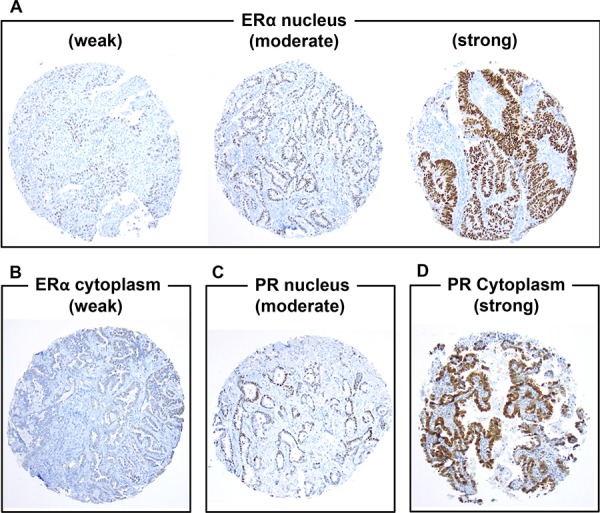
We reviewed patients with pathologic stage I resected lung adenocarcinoma. Tumors were classified according to the IASLC/ATS/ERS classification. Immunostaining of ERα and PR was performed using tissue microarrays (n = 913). Immunostaining of CD3+ and forkhead box P3 (FoxP3)+ lymphocyte infiltration, interleukin-7 receptor (IL-7R), and IL-12Rβ2 were performed. Cumulative incidence of recurrence (CIR) analysis was used to estimate probability of recurrence.
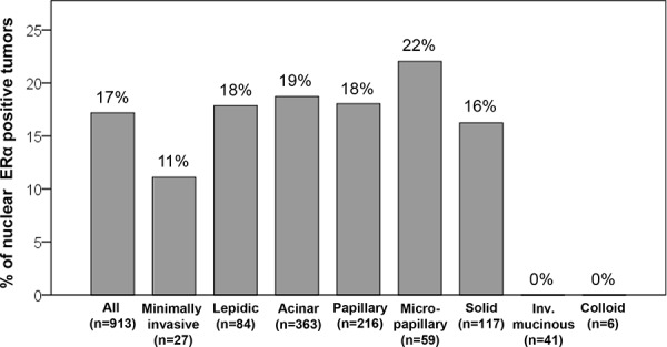
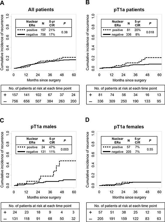
Nuclear ERα expression was observed in 157 (17%) patients and presented more frequently in females (P = 0.038) and smaller tumors (P = 0.019). Nuclear ERα expression was not identified in mucinous tumors. In pT1a patients, 5-year CIR of patients with ERα-positive tumors was significantly higher (5-year CIR, 20%) than those with ERα-negative tumors (8%; P = 0.018). This difference was statistically significant in males (P = 0.003) but not females (P = 0.55). On multivariate analysis, nuclear ERα expression was an independent predictor of recurrence (hazard ratio = 2.27; P = 0.030). In pT1a patients, nuclear ERα expression positively correlated with tumoral FoxP3+ lymphocytes (P < 0.001), FoxP3/CD3 index (P < 0.001), and IL-7R (P = 0.022).
Nuclear ERα expression is an independent predictor of recurrence in pT1a lung adenocarcinomas and correlates with poor prognostic immune microenvironments.
雌激素受体-α(ERα)和孕激素受体(PR)的肿瘤生物学特性已在乳腺癌中得到研究。然而,其在肺癌中的临床影响仍存在争议。在我们的研究中,我们调查ERα和PR表达是否可预测疾病复发以及是否与I期肺腺癌的免疫因素相关。
我们回顾了经手术切除的病理I期肺腺癌患者。肿瘤根据国际肺癌研究协会(IASLC)/美国胸科学会(ATS)/欧洲呼吸学会(ERS)分类进行分类。使用组织微阵列(n = 913)对ERα和PR进行免疫染色。对CD3 +和叉头框P3(FoxP3)+淋巴细胞浸润、白细胞介素-7受体(IL-7R)和IL-12Rβ2进行免疫染色。采用复发累积发生率(CIR)分析来估计复发概率。
157例(17%)患者观察到核ERα表达,在女性患者中更常见(P = 0.038),且在较小肿瘤中更常见(P = 0.019)。黏液性肿瘤中未发现核ERα表达。在pT1a患者中,ERα阳性肿瘤患者的5年CIR显著高于ERα阴性肿瘤患者(5年CIR,20%对8%;P = 0.018)。这种差异在男性中具有统计学意义(P = 0.003),但在女性中无统计学意义(P = 0.55)。多因素分析显示,核ERα表达是复发的独立预测因素(风险比 = 2.27;P = 0.030)。在pT1a患者中,核ERα表达与肿瘤内FoxP3 +淋巴细胞(P < 0.001)、FoxP3/CD3指数(P < 0.001)和IL-7R(P = 0.022)呈正相关。
核ERα表达是pT1a肺腺癌复发的独立预测因素,且与不良预后的免疫微环境相关。