Boyer Richard B, Kelm Nathaniel D, Riley D Colton, Sexton Kevin W, Pollins Alonda C, Shack R Bruce, Dortch Richard D, Nanney Lillian B, Does Mark D, Thayer Wesley P
Departments of 1 Biomedical Engineering and.
Plastic Surgery, Vanderbilt University Medical Center;
Neurosurg Focus. 2015 Sep;39(3):E9. doi: 10.3171/2015.6.FOCUS1590.
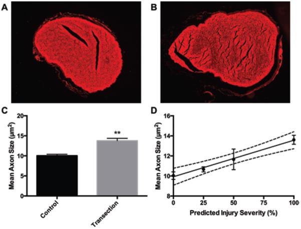
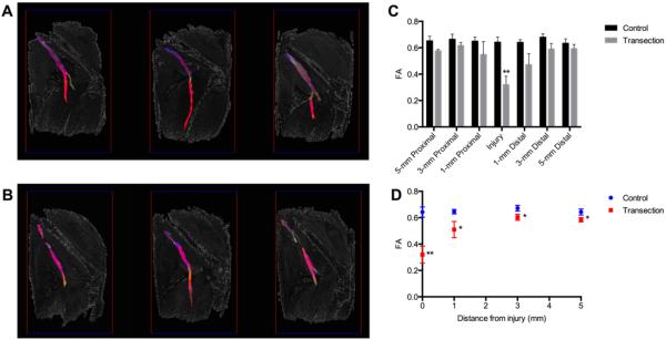
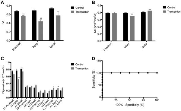
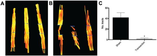
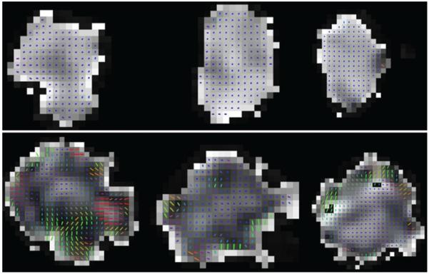
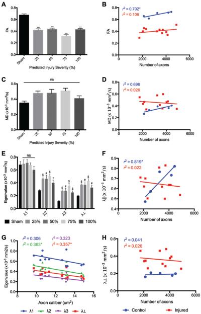
Diagnosis and management of peripheral nerve injury is complicated by the inability to assess microstructural features of injured nerve fibers via clinical examination and electrophysiology. Diffusion tensor imaging (DTI) has been shown to accurately detect nerve injury and regeneration in crush models of peripheral nerve injury, but no prior studies have been conducted on nerve transection, a surgical emergency that can lead to permanent weakness or paralysis. Acute sciatic nerve injuries were performed microsurgically to produce multiple grades of nerve transection in rats that were harvested 1 hour after surgery. High-resolution diffusion tensor images from ex vivo sciatic nerves were obtained using diffusion-weighted spin-echo acquisitions at 4.7 T. Fractional anisotropy was significantly reduced at the injury sites of transected rats compared with sham rats. Additionally, minor eigenvalues and radial diffusivity were profoundly elevated at all injury sites and were negatively correlated to the degree of injury. Diffusion tensor tractography showed discontinuities at all injury sites and significantly reduced continuous tract counts. These findings demonstrate that high-resolution DTI is a promising tool for acute diagnosis and grading of traumatic peripheral nerve injuries.
由于无法通过临床检查和电生理学评估受损神经纤维的微观结构特征,周围神经损伤的诊断和管理变得复杂。扩散张量成像(DTI)已被证明能在周围神经损伤的挤压模型中准确检测神经损伤和再生,但此前尚未有关于神经横断伤的研究,而神经横断伤是一种可导致永久性无力或瘫痪的外科急症。通过显微外科手术造成大鼠急性坐骨神经损伤,产生多个等级的神经横断伤,并在术后1小时处死大鼠。使用4.7 T的扩散加权自旋回波采集技术,获得离体坐骨神经的高分辨率扩散张量图像。与假手术组大鼠相比,横断伤大鼠损伤部位的分数各向异性显著降低。此外,所有损伤部位的最小本征值和径向扩散率均显著升高,且与损伤程度呈负相关。扩散张量纤维束成像显示所有损伤部位均存在纤维束中断,且连续纤维束计数显著减少。这些发现表明,高分辨率DTI是一种用于创伤性周围神经损伤急性诊断和分级的有前景的工具。