Vanderbilt University Medical Center, Department Radiology and Radiological Sciences, Nashville, TN, United States of America; Vanderbilt University Medical Center, Institute of Imaging Science, Nashville, TN, United States of America.
Vanderbilt University Medical Center, Department of Plastic Surgery, Nashville, TN, United States of America.
Magn Reson Imaging. 2021 Nov;83:96-106. doi: 10.1016/j.mri.2021.08.006. Epub 2021 Aug 14.
Primary repair of peripheral nerves is recommended following transection; however, patient management following repair is challenged by a lack of biomarkers to nerve regeneration. Previous studies have demonstrated that diffusion magnetic resonance imaging (MRI) may provide viable biomarkers of nerve regeneration in injury models; though, these methods have not been systematically evaluated in graded partial transections and repairs.
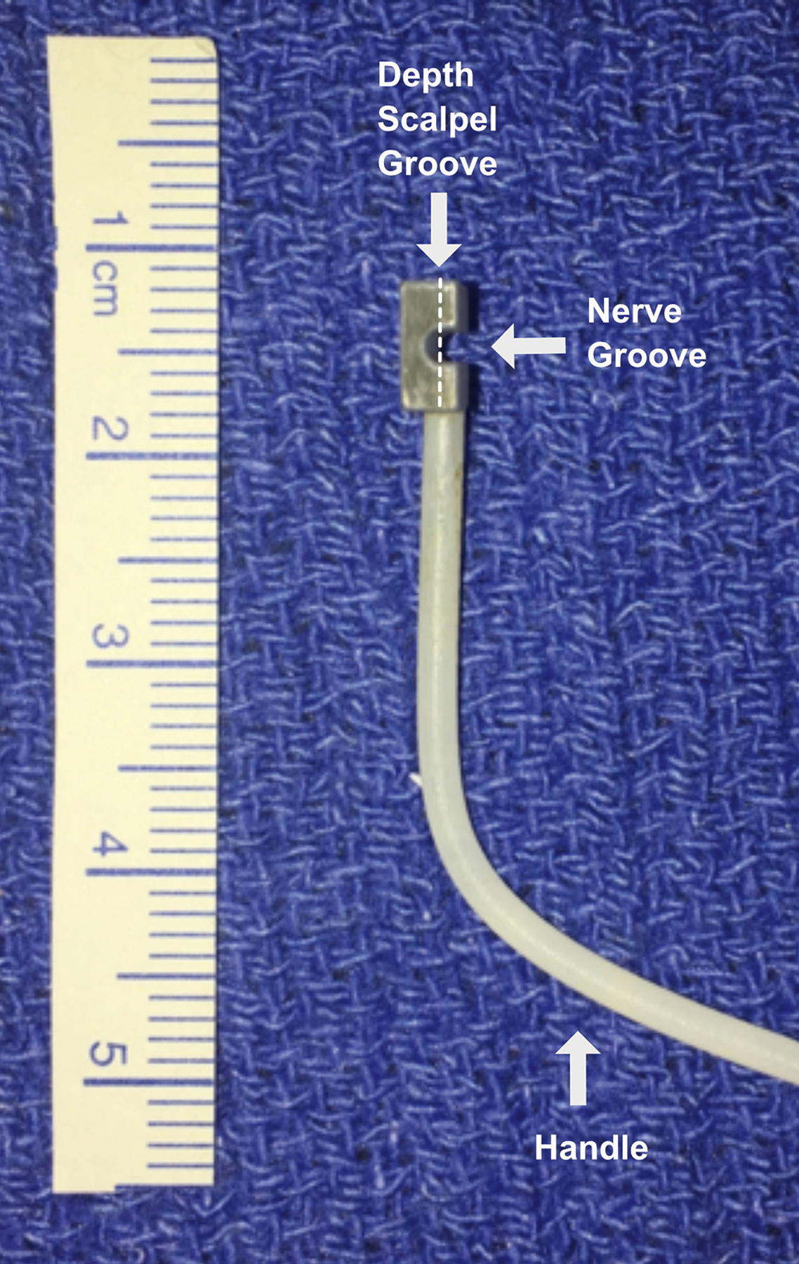
Ex vivo diffusion MRI was performed in fixed rat sciatic nerve samples 4 or 12 weeks following partial nerve transection and repair (25% cut = 12, 50% cut = 12 and 75% cut = 11), crush injuries (n = 12), and sham surgeries (n = 9). Behavioral testing and histologic evaluation were performed in the same animals and nerve samples for comparison.
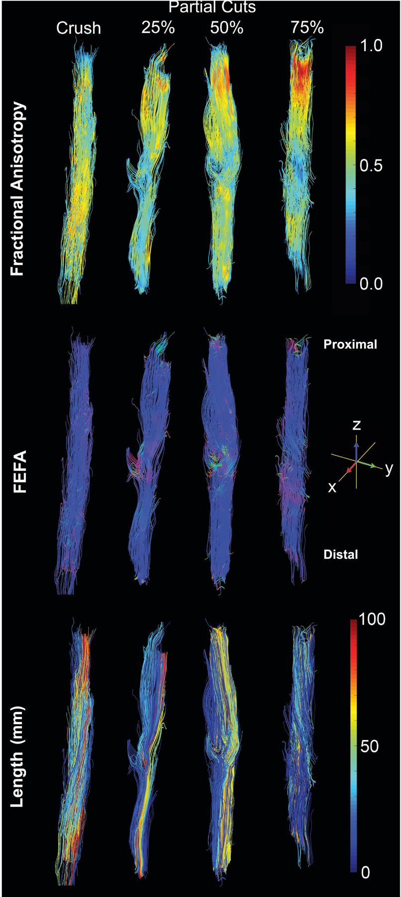
Diffusion tractography provided visual characterizations of nerve damage and recovery consistent with the expected degree of injury within each cohort. In addition, quantitative indices from diffusion MRI correlated with both histological and behavioral evaluations, the latter of indicated full recovery for sham and crush nerves and limited recovery in all partially transected/repaired nerves. Nerve recovery between 4 and 12 weeks was statistically significant in partial transections 50% and 75% depth cuts (p = 0.043 and p = 0.022) but not for 25% transections.
Our findings suggest that DTI can i) distinguish different degrees of partial nerve transection following surgical repair and ii) map spatially heterogeneous nerve recovery (e.g., due to collateral sprouting) from 4 to 12 weeks in partially transected nerves.
神经离断后推荐行一期修复;然而,修复后患者管理面临缺乏神经再生的生物标志物的挑战。先前的研究表明,弥散磁共振成像(MRI)可能为损伤模型中的神经再生提供可行的生物标志物;尽管这些方法尚未在分级部分横断和修复中得到系统评估。
对部分神经横断和修复(25%切断=12 根、50%切断=12 根和 75%切断=11 根)、挤压伤(n=12)和假手术(n=9)后的固定大鼠坐骨神经样本进行离体弥散 MRI 检查。在相同动物和神经样本中进行行为学测试和组织学评估进行比较。
弥散轨迹提供了与每个队列中预期损伤程度一致的神经损伤和恢复的直观特征。此外,弥散 MRI 的定量指标与组织学和行为学评估相关,后者表明假手术和挤压伤神经完全恢复,所有部分横断/修复神经恢复有限。50%和 75%深度横断的部分横断神经在 4 至 12 周时神经恢复具有统计学意义(p=0.043 和 p=0.022),但 25%横断神经无统计学意义。
我们的发现表明,DTI 可以 i)区分手术后不同程度的部分神经横断,ii)在部分横断神经中从 4 周到 12 周映射空间异质的神经恢复(例如,由于侧支发芽)。