Eichorst Stephanie A, Strasser Florian, Woyke Tanja, Schintlmeister Arno, Wagner Michael, Woebken Dagmar
Division of Microbial Ecology, Department of Microbiology and Ecosystem Science, Research network 'Chemistry meets Microbiology', University of Vienna, Vienna 1090 Austria.
DOE Joint Genome Institute, Walnut Creek, CA 94598, USA.
FEMS Microbiol Ecol. 2015 Oct;91(10). doi: 10.1093/femsec/fiv106. Epub 2015 Aug 30.
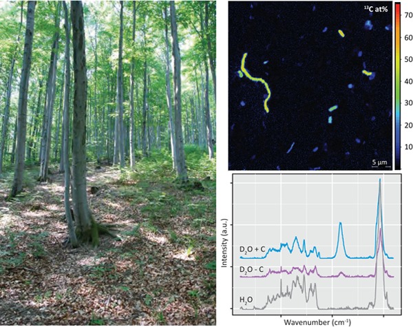
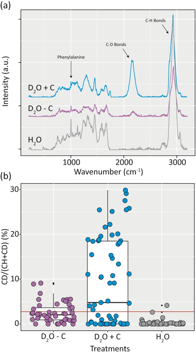
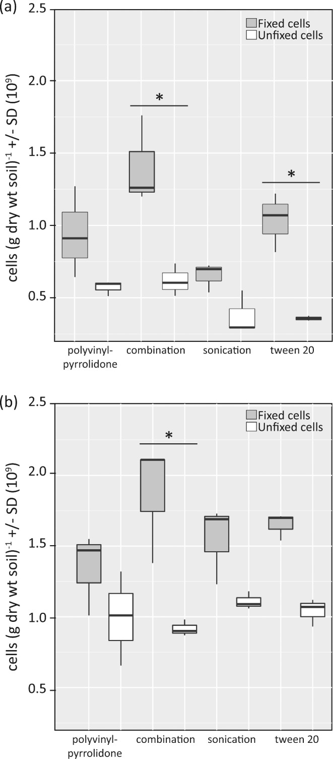
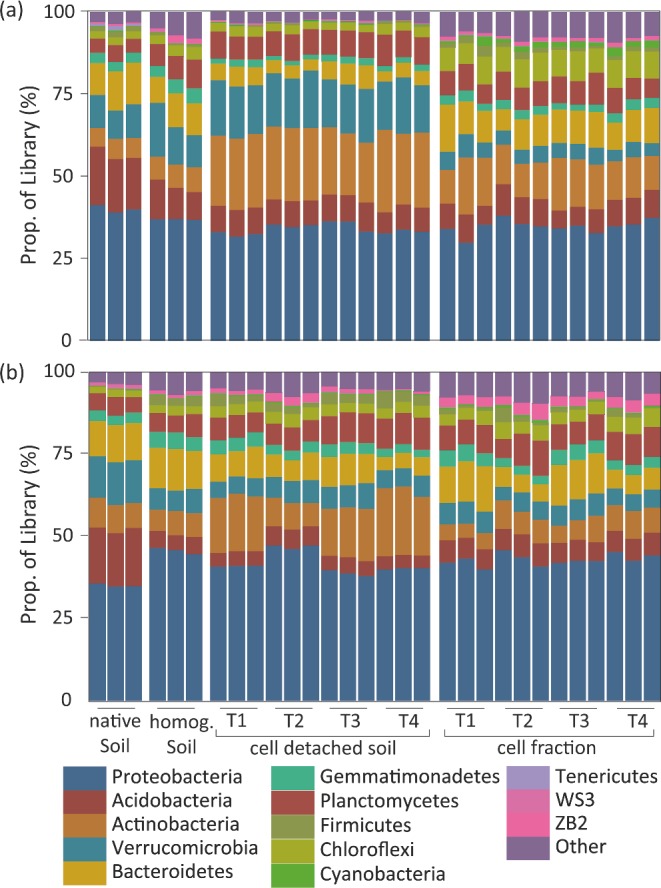
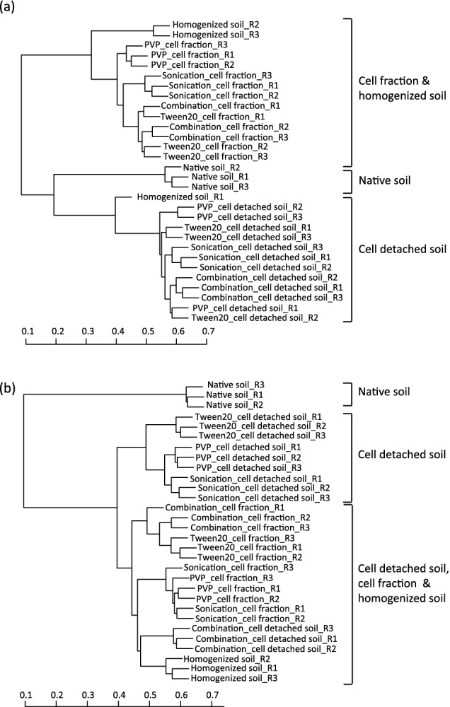
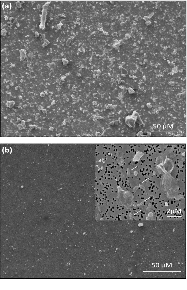
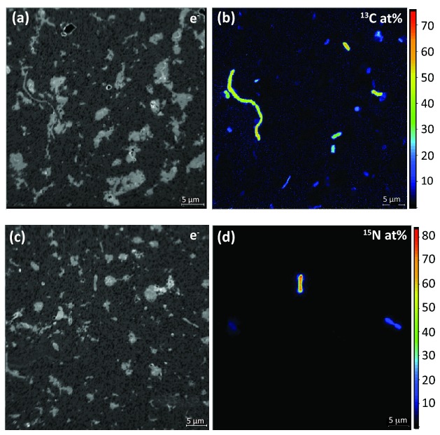
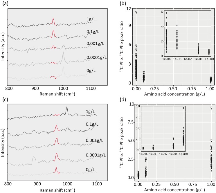
The combined approach of incubating environmental samples with stable isotope-labeled substrates followed by single-cell analyses through high-resolution secondary ion mass spectrometry (NanoSIMS) or Raman microspectroscopy provides insights into the in situ function of microorganisms. This approach has found limited application in soils presumably due to the dispersal of microbial cells in a large background of particles. We developed a pipeline for the efficient preparation of cell extracts from soils for subsequent single-cell methods by combining cell detachment with separation of cells and soil particles followed by cell concentration. The procedure was evaluated by examining its influence on cell recoveries and microbial community composition across two soils. This approach generated a cell fraction with considerably reduced soil particle load and of sufficient small size to allow single-cell analysis by NanoSIMS, as shown when detecting active N2-fixing and cellulose-responsive microorganisms via (15)N2 and (13)C-UL-cellulose incubations, respectively. The same procedure was also applicable for Raman microspectroscopic analyses of soil microorganisms, assessed via microcosm incubations with a (13)C-labeled carbon source and deuterium oxide (D2O, a general activity marker). The described sample preparation procedure enables single-cell analysis of soil microorganisms using NanoSIMS and Raman microspectroscopy, but should also facilitate single-cell sorting and sequencing.
将环境样品与稳定同位素标记的底物一起孵育,随后通过高分辨率二次离子质谱(纳米二次离子质谱仪)或拉曼光谱进行单细胞分析的联合方法,能够深入了解微生物的原位功能。这种方法在土壤中的应用有限,可能是由于微生物细胞分散在大量的颗粒背景中。我们开发了一种流程,通过将细胞分离与细胞和土壤颗粒的分离相结合,随后进行细胞浓缩,从土壤中高效制备细胞提取物,用于后续的单细胞方法。通过考察该流程对两种土壤中细胞回收率和微生物群落组成的影响来对其进行评估。这种方法产生了一个土壤颗粒负载显著降低且尺寸足够小的细胞组分,以便通过纳米二次离子质谱仪进行单细胞分析,这在分别通过(15)N2和(13)C - UL - 纤维素孵育检测活跃的固氮微生物和纤维素响应微生物时得到了证明。相同的流程也适用于对土壤微生物进行拉曼光谱分析,通过与(13)C标记的碳源和氧化氘(D2O,一种通用的活性标记物)进行微观培养来评估。所描述的样品制备流程能够使用纳米二次离子质谱仪和拉曼光谱对土壤微生物进行单细胞分析,而且还应便于单细胞分选和测序。