Zhang Bo, Cowden Daniel, Zhang Fan, Yuan Jue, Siedlak Sandra, Abouelsaad Mai, Zeng Liang, Zhou Xuefeng, O'Toole John, Das Alvin S, Kofskey Diane, Warren Miriam, Bian Zehua, Cui Yuqi, Tan Tao, Kresak Adam, Wyza Robert E, Petersen Robert B, Wang Gong-Xian, Kong Qingzhong, Wang Xinglong, Sedor John, Zhu Xiongwei, Zhu Hua, Zou Wen-Quan
Institute of Organ Transplantation, Tongji Hospital, Tongji Medical College, Huazhong University of Science and Technology, Wuhan, HuBei, The People's Republic of China; Department of Surgery, Davis Heart and Lung Research Institute, The Ohio State University, Columbus, Ohio, United States of America; Key Laboratory of Ministry of Health and Key Laboratory of Ministry of Education, Wuhan, HuBei, The People's Republic of China.
Department of Pathology, Case Western Reserve University/University Hospitals Case Medical Center, Cleveland, Ohio, United States of America.
PLoS One. 2015 Sep 1;10(9):e0136923. doi: 10.1371/journal.pone.0136923. eCollection 2015.
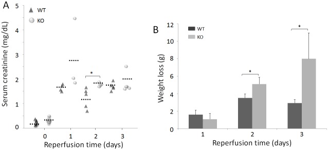
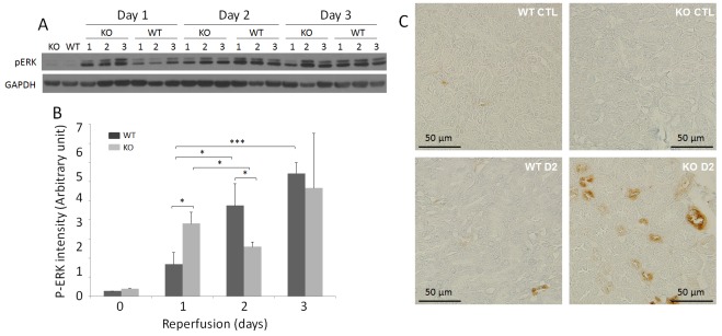
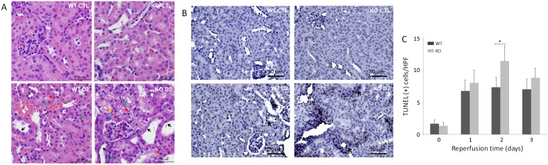
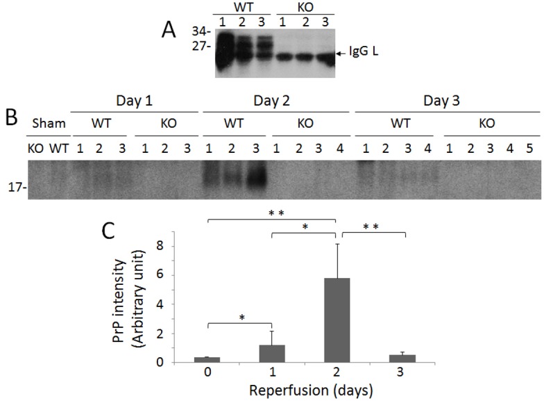
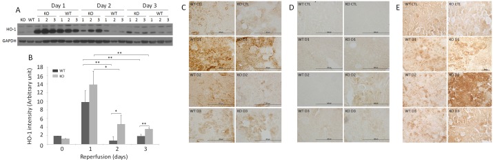
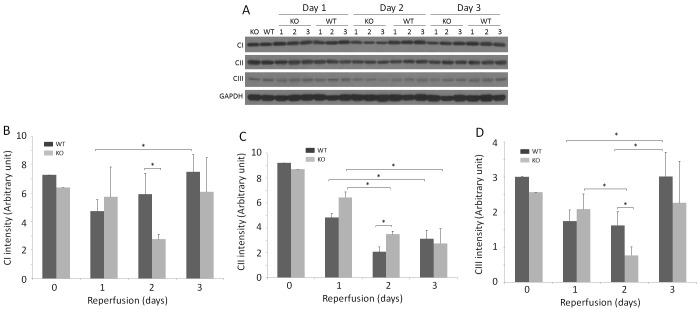
The cellular prion protein (PrPC), a protein most noted for its link to prion diseases, has been found to play a protective role in ischemic brain injury. To investigate the role of PrPC in the kidney, an organ highly prone to ischemia/reperfusion (IR) injury, we examined wild-type (WT) and PrPC knockout (KO) mice that were subjected to 30-min of renal ischemia followed by 1, 2, or 3 days of reperfusion. Renal dysfunction and structural damage was more severe in KO than in WT mice. While PrP was undetectable in KO kidneys, Western blotting revealed an increase in PrP in IR-injured WT kidneys compared to sham-treated kidneys. Compared to WT, KO kidneys exhibited increases in oxidative stress markers heme oxygenase-1, nitrotyrosine, and Nε-(carboxymethyl)lysine, and decreases in mitochondrial complexes I and III. Notably, phosphorylated extracellular signal-regulated kinase (pERK) staining was predominantly observed in tubular cells from KO mice following 2 days of reperfusion, a time at which significant differences in renal dysfunction, histological changes, oxidative stress, and mitochondrial complexes between WT and KO mice were observed. Our study provides the first evidence that PrPC may play a protective role in renal IR injury, likely through its effects on mitochondria and ERK signaling pathways.
细胞朊蛋白(PrPC),一种最因与朊病毒疾病相关而闻名的蛋白质,已被发现对缺血性脑损伤具有保护作用。为了研究PrPC在肾脏(一个极易发生缺血/再灌注(IR)损伤的器官)中的作用,我们检查了野生型(WT)和PrPC基因敲除(KO)小鼠,这些小鼠经历了30分钟的肾脏缺血,随后再灌注1、2或3天。KO小鼠的肾功能障碍和结构损伤比WT小鼠更严重。虽然在KO小鼠的肾脏中检测不到PrP,但蛋白质印迹法显示,与假手术处理的肾脏相比,IR损伤的WT小鼠肾脏中PrP增加。与WT小鼠相比,KO小鼠的肾脏中氧化应激标志物血红素加氧酶-1、硝基酪氨酸和Nε-(羧甲基)赖氨酸增加,线粒体复合物I和III减少。值得注意的是,再灌注2天后,在KO小鼠的肾小管细胞中主要观察到磷酸化细胞外信号调节激酶(pERK)染色,此时在WT和KO小鼠之间观察到肾功能障碍、组织学变化、氧化应激和线粒体复合物存在显著差异。我们的研究提供了首个证据,表明PrPC可能在肾脏IR损伤中发挥保护作用,可能是通过其对线粒体和ERK信号通路的影响。