Pretorius A J, Zhou Y, Ruddle R A
BMC Bioinformatics. 2015;16 Suppl 11(Suppl 11):S9. doi: 10.1186/1471-2105-16-S11-S9. Epub 2015 Aug 13.
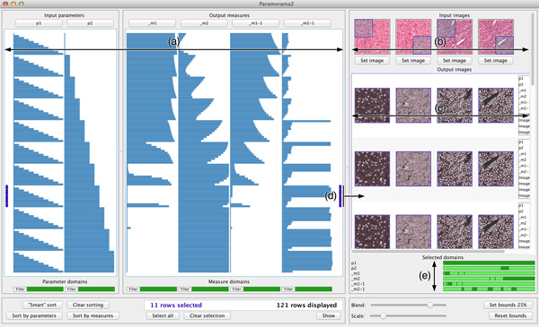
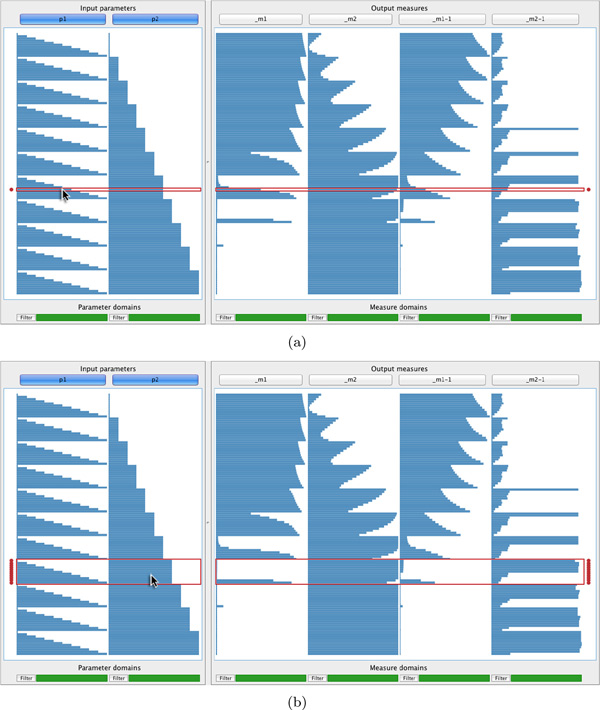
Biomedical image processing methods require users to optimise input parameters to ensure high-quality output. This presents two challenges. First, it is difficult to optimise multiple input parameters for multiple input images. Second, it is difficult to achieve an understanding of underlying algorithms, in particular, relationships between input and output.
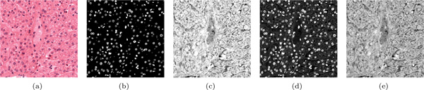
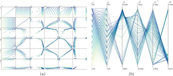
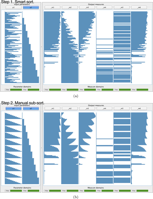
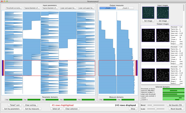
We present a visualisation method that transforms users' ability to understand algorithm behaviour by integrating input and output, and by supporting exploration of their relationships. We discuss its application to a colour deconvolution technique for stained histology images and show how it enabled a domain expert to identify suitable parameter values for the deconvolution of two types of images, and metrics to quantify deconvolution performance. It also enabled a breakthrough in understanding by invalidating an underlying assumption about the algorithm.
The visualisation method presented here provides analysis capability for multiple inputs and outputs in biomedical image processing that is not supported by previous analysis software. The analysis supported by our method is not feasible with conventional trial-and-error approaches.
生物医学图像处理方法要求用户优化输入参数以确保高质量输出。这带来了两个挑战。首先,为多个输入图像优化多个输入参数很困难。其次,难以理解底层算法,尤其是输入与输出之间的关系。
我们提出了一种可视化方法,通过整合输入和输出,并支持对它们之间关系的探索,来转变用户理解算法行为的能力。我们讨论了其在用于染色组织学图像的颜色反卷积技术中的应用,并展示了它如何使领域专家能够为两种类型图像的反卷积确定合适的参数值,以及量化反卷积性能的指标。它还通过推翻关于该算法的一个基本假设实现了理解上的突破。
本文提出的可视化方法为生物医学图像处理中的多个输入和输出提供了分析能力,这是以前的分析软件所不具备的。我们的方法所支持的分析用传统的试错方法是不可行的。