• 文献检索
  • 文档翻译
  • 深度研究
  • 学术资讯
  • Suppr Zotero 插件Zotero 插件
  • 邀请有礼
  • 套餐&价格
  • 历史记录
应用&插件
Suppr Zotero 插件Zotero 插件浏览器插件Mac 客户端Windows 客户端微信小程序
定价
高级版会员购买积分包购买API积分包
服务
文献检索文档翻译深度研究API 文档MCP 服务
关于我们
关于 Suppr公司介绍联系我们用户协议隐私条款
关注我们

Suppr 超能文献

核心技术专利:CN118964589B侵权必究
粤ICP备2023148730 号-1Suppr @ 2026

文献检索

告别复杂PubMed语法,用中文像聊天一样搜索,搜遍4000万医学文献。AI智能推荐,让科研检索更轻松。

立即免费搜索

文件翻译

保留排版,准确专业,支持PDF/Word/PPT等文件格式,支持 12+语言互译。

免费翻译文档

深度研究

AI帮你快速写综述,25分钟生成高质量综述,智能提取关键信息,辅助科研写作。

立即免费体验

远场无标记超分辨率成像的新方法。

Novel approach for label free super-resolution imaging in far field.

作者信息

Alexandrov Sergey A, McGrath James, Subhash Hrebesh, Boccafoschi Francesca, Giannini Cinzia, Leahy Martin

机构信息

Tissue Optics &Microcirculation Imaging Group, School of Physics, National University of Ireland, Galway, Ireland.

Department of Health Sciences, University of Piemonte Orientale "A. Avogadro", 28100 Novara, Italy.

出版信息

Sci Rep. 2015 Sep 3;5:13274. doi: 10.1038/srep13274.

DOI:10.1038/srep13274
PMID:26333595
原文链接:https://pmc.ncbi.nlm.nih.gov/articles/PMC4558609/
Abstract

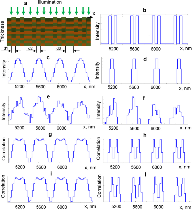
Progress in the emerging areas of science and technology, such as bio- and nano-technologies, depends on development of corresponding techniques for imaging and probing the structures with high resolution. Recently, the far field diffraction resolution limit in the optical range has been circumvented and different methods of super-resolution optical microscopy have been developed. The importance of this breakthrough achievement has been recognized by Nobel Prize for Chemistry in 2014. However, the fluorescence based super-resolution techniques only function with fluorescent molecules (most of which are toxic and can destroy or lead to artificial results in living biological objects) and suffer from photobleaching. Here we show a new way to break the diffraction resolution limit, which is based on nano-sensitivity to internal structure. Instead of conventional image formation as 2D intensity distribution, in our approach images are formed as a result of comparison of the axial spatial frequency profiles, reconstructed for each image point. The proposed approach dramatically increases the lateral resolution even in presence of noise and allows objects to be imaged in their natural state, without any labels.

摘要

诸如生物和纳米技术等新兴科技领域的进展,取决于开发相应的高分辨率成像和探测结构的技术。最近,光学范围内的远场衍射分辨率极限已被突破,并且已经开发出不同的超分辨率光学显微镜方法。这一突破性成就的重要性已在2014年获得诺贝尔化学奖认可。然而,基于荧光的超分辨率技术仅对荧光分子起作用(其中大多数有毒,会破坏活的生物物体或导致人为结果),并且存在光漂白问题。在此,我们展示了一种突破衍射分辨率极限的新方法,该方法基于对内部结构的纳米敏感性。与传统的作为二维强度分布的图像形成方式不同,在我们的方法中,图像是通过比较为每个图像点重建的轴向空间频率分布而形成的。所提出的方法即使在存在噪声的情况下也能显著提高横向分辨率,并允许在物体处于自然状态下成像,无需任何标记。

https://cdn.ncbi.nlm.nih.gov/pmc/blobs/e628/4558609/ebc841dfea1a/srep13274-f5.jpg
https://cdn.ncbi.nlm.nih.gov/pmc/blobs/e628/4558609/1c3801766bc9/srep13274-f1.jpg
https://cdn.ncbi.nlm.nih.gov/pmc/blobs/e628/4558609/e482241d1764/srep13274-f2.jpg
https://cdn.ncbi.nlm.nih.gov/pmc/blobs/e628/4558609/969d267c7ed6/srep13274-f3.jpg
https://cdn.ncbi.nlm.nih.gov/pmc/blobs/e628/4558609/b99437939b40/srep13274-f4.jpg
https://cdn.ncbi.nlm.nih.gov/pmc/blobs/e628/4558609/ebc841dfea1a/srep13274-f5.jpg

相似文献

1
Novel approach for label free super-resolution imaging in far field.远场无标记超分辨率成像的新方法。
Sci Rep. 2015 Sep 3;5:13274. doi: 10.1038/srep13274.
2
Increasing microscopy resolution with photobleaching and intensity cumulant analysis.通过光漂白和强度累积分析提高显微镜分辨率。
Microsc Res Tech. 2015 Nov;78(11):958-68. doi: 10.1002/jemt.22559. Epub 2015 Aug 17.
3
Recent advances in super-resolution fluorescence imaging and its applications in biology.超分辨率荧光成像技术的最新进展及其在生物学中的应用。
J Genet Genomics. 2013 Dec 20;40(12):583-95. doi: 10.1016/j.jgg.2013.11.003. Epub 2013 Nov 23.
4
Super-resolution nonlinear photothermal microscopy.超分辨率非线性光热显微镜。
Small. 2014 Jan 15;10(1):135-42. doi: 10.1002/smll.201300024. Epub 2013 Jul 17.
5
Label-free ultra-sensitive visualization of structure below the diffraction resolution limit.低于衍射分辨率极限的结构的无标记超灵敏可视化。
J Biophotonics. 2018 Jul;11(7):e201700385. doi: 10.1002/jbio.201700385. Epub 2018 May 9.
6
Image scanning microscopy: an overview.图像扫描显微镜:概述。
J Microsc. 2017 May;266(2):221-228. doi: 10.1111/jmi.12534. Epub 2017 Mar 1.
7
Development of a reversibly switchable fluorescent protein for super-resolution optical fluctuation imaging (SOFI).用于超分辨率光学波动成像(SOFI)的可反向切换荧光蛋白的开发。
ACS Nano. 2015 Mar 24;9(3):2659-67. doi: 10.1021/nn5064387. Epub 2015 Feb 23.
8
Nanoparticles as Nonfluorescent Analogues of Fluorophores for Optical Nanoscopy.纳米粒子作为荧光团的非荧光类似物在光学纳米技术中的应用。
ACS Nano. 2015 Jun 23;9(6):6196-205. doi: 10.1021/acsnano.5b01503. Epub 2015 May 15.
9
Roadmap on Label-Free Super-Resolution Imaging.无标记超分辨率成像路线图
Laser Photon Rev. 2023 Dec;17(12). doi: 10.1002/lpor.202200029. Epub 2023 Oct 30.
10
Sparsity-based super-resolution microscopy from correlation information.基于相关性信息的稀疏超分辨率显微镜技术。
Opt Express. 2018 Jul 9;26(14):18238-18269. doi: 10.1364/OE.26.018238.

引用本文的文献

1
Skin cancer margin detection using nanosensitive optical coherence tomography and a comparative study with confocal microscopy.使用纳米敏感光学相干断层扫描技术进行皮肤癌边缘检测以及与共聚焦显微镜的对比研究。
Biomed Opt Express. 2022 Oct 7;13(11):5654-5666. doi: 10.1364/BOE.474334. eCollection 2022 Nov 1.
2
Accessing depth-resolved high spatial frequency content from the optical coherence tomography signal.从光相干断层扫描信号中获取深度分辨的高空间频率内容。
Sci Rep. 2021 Aug 24;11(1):17123. doi: 10.1038/s41598-021-96619-7.
3
Nanosensitive optical coherence tomography to assess wound healing within the cornea.

本文引用的文献

1
Three-dimensional super-resolution and localization of dense clusters of single molecules.单分子密集簇的三维超分辨率与定位
Sci Rep. 2014 Jun 23;4:5388. doi: 10.1038/srep05388.
2
Single-molecule reconstruction of oligonucleotide secondary structure by atomic force microscopy.原子力显微镜中单链寡核苷酸二级结构的重构。
Small. 2014 Aug 27;10(16):3257-61. doi: 10.1002/smll.201400265. Epub 2014 Apr 17.
3
Nano-sensitive optical coherence tomography.纳米敏感染光相干断层扫描技术。
用于评估角膜内伤口愈合情况的纳米敏感光学相干断层扫描技术。
Biomed Opt Express. 2020 Jun 2;11(7):3407-3422. doi: 10.1364/BOE.389342. eCollection 2020 Jul 1.
4
Review of interferometric spectroscopy of scattered light for the quantification of subdiffractional structure of biomaterials.用于定量生物材料亚衍射结构的漫散射光干涉光谱学综述。
J Biomed Opt. 2017 Mar 1;22(3):30901. doi: 10.1117/1.JBO.22.3.030901.
5
Multiscale dispersion-state characterization of nanocomposites using optical coherence tomography.利用光学相干断层扫描对纳米复合材料进行多尺度分散状态表征。
Sci Rep. 2016 Aug 25;6:31733. doi: 10.1038/srep31733.
6
Functional imaging for regenerative medicine.再生医学的功能成像
Stem Cell Res Ther. 2016 Apr 19;7(1):57. doi: 10.1186/s13287-016-0315-2.
Nanoscale. 2014 Apr 7;6(7):3545-9. doi: 10.1039/c3nr06132a.
4
Coherent total internal reflection dark-field microscopy: label-free imaging beyond the diffraction limit.相干全内反射暗场显微镜:超越衍射极限的无标记成像。
Opt Lett. 2013 Oct 15;38(20):4066-9. doi: 10.1364/OL.38.004066.
5
Tomographic imaging via spectral encoding of spatial frequency.通过空间频率的光谱编码进行断层成像。
Opt Express. 2013 Mar 25;21(6):7488-504. doi: 10.1364/OE.21.007488.
6
Breaking the diffraction barrier using fluorescence emission difference microscopy.利用荧光发射差显微镜突破衍射障碍。
Sci Rep. 2013;3:1441. doi: 10.1038/srep01441.
7
A framework for far-field infrared absorption microscopy beyond the diffraction limit.一种超越衍射极限的远场红外吸收显微镜框架。
Opt Express. 2012 Dec 31;20(28):29694-704. doi: 10.1364/OE.20.029694.
8
Simple super-resolution live-cell imaging based on diffusion-assisted Förster resonance energy transfer.基于扩散辅助Förster 共振能量转移的简单超分辨率活细胞成像。
Sci Rep. 2013;3:1208. doi: 10.1038/srep01208. Epub 2013 Feb 4.
9
Spectral encoding of spatial frequency approach for characterization of nanoscale structures.用于纳米级结构表征的空间频率方法的光谱编码
Appl Phys Lett. 2012 Jul 16;101(3):33702. doi: 10.1063/1.4737209. Epub 2012 Jul 17.
10
Spectral contrast imaging microscopy.光谱对比成像显微镜。
Opt Lett. 2011 Sep 1;36(17):3323-5. doi: 10.1364/OL.36.003323.