Department of Electrical and Computer Engineering, Drexel University, Philadelphia, PA 19104, USA.
Neural Stem Cell Institute, Rensselaer, NY 12144, USA.
Stem Cell Reports. 2015 Oct 13;5(4):609-20. doi: 10.1016/j.stemcr.2015.08.002. Epub 2015 Sep 3.
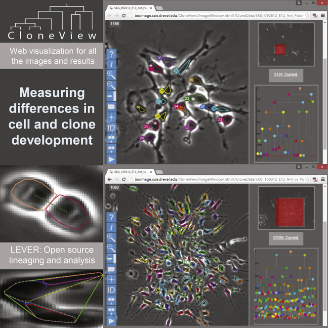
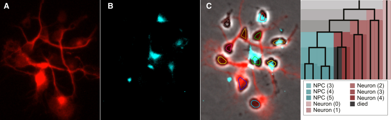
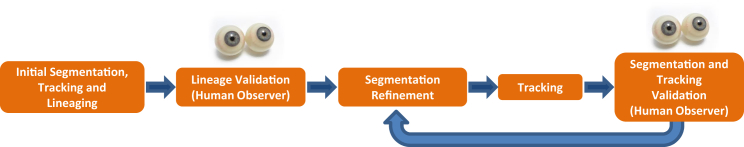
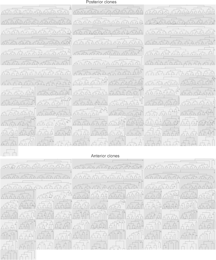
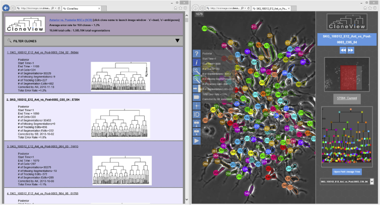
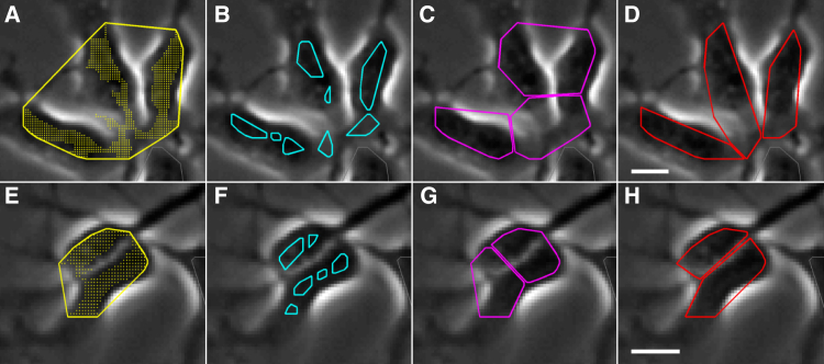
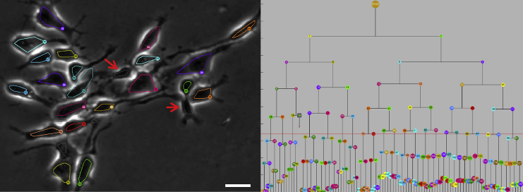
Time-lapse microscopy can capture patterns of development through multiple divisions for an entire clone of proliferating cells. Images are taken every few minutes over many days, generating data too vast to process completely by hand. Computational analysis of this data can benefit from occasional human guidance. Here we combine improved automated algorithms with minimized human validation to produce fully corrected segmentation, tracking, and lineaging results with dramatic reduction in effort. A web-based viewer provides access to data and results. The improved approach allows efficient analysis of large numbers of clones. Using this method, we studied populations of progenitor cells derived from the anterior and posterior embryonic mouse cerebral cortex, each growing in a standardized culture environment. Progenitors from the anterior cortex were smaller, less motile, and produced smaller clones compared to those from the posterior cortex, demonstrating cell-intrinsic differences that may contribute to the areal organization of the cerebral cortex.
延时显微镜可以通过对整个增殖细胞克隆进行多次分裂来捕获发育模式。每隔几分钟拍摄一次图像,持续数天,生成的数据过于庞大,无法完全手动处理。对这些数据的计算分析可以受益于偶尔的人工指导。在这里,我们将改进的自动化算法与最小化的人工验证相结合,以产生完全校正的分割、跟踪和谱系结果,从而大大减少工作量。一个基于网络的查看器提供了对数据和结果的访问。改进的方法允许对大量克隆进行高效分析。使用这种方法,我们研究了来自胚胎小鼠大脑前脑和后脑的祖细胞群体,每个群体都在标准化的培养环境中生长。与来自后脑的祖细胞相比,前脑的祖细胞更小、运动能力更差,并且产生的克隆更小,这表明细胞内在的差异可能有助于大脑皮层的区域组织。