Pourfarhangi Kamyar Esmaeili, De La Hoz Edgar Cardenas, Cohen Andrew R, Gligorijevic Bojana
Bioengineering department, College of Engineering, Temple University, Philadelphia, Pennsylvania 19122, USA.
Department of Electrical and Computer Engineering, College of Engineering, Drexel University, Philadelphia, Pennsylvania 19104, USA.
APL Bioeng. 2018 Sep;2(3). doi: 10.1063/1.5026419. Epub 2018 May 30.
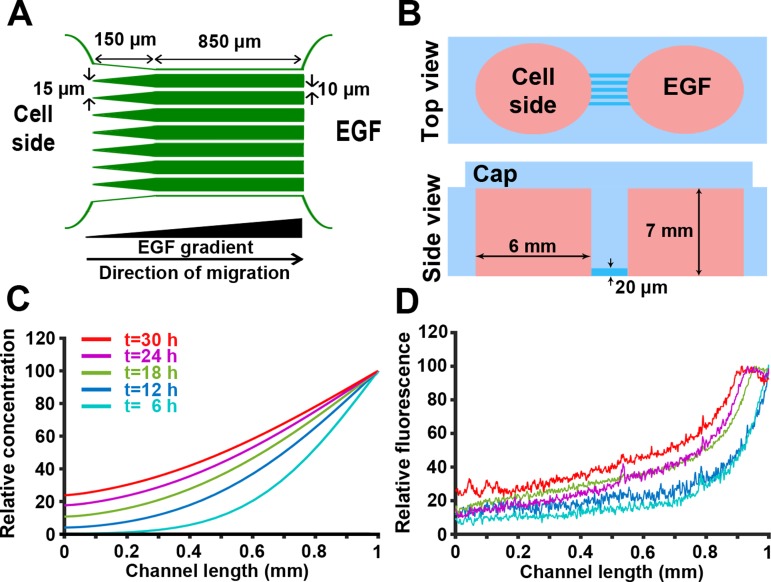
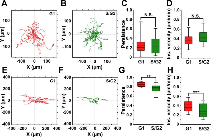
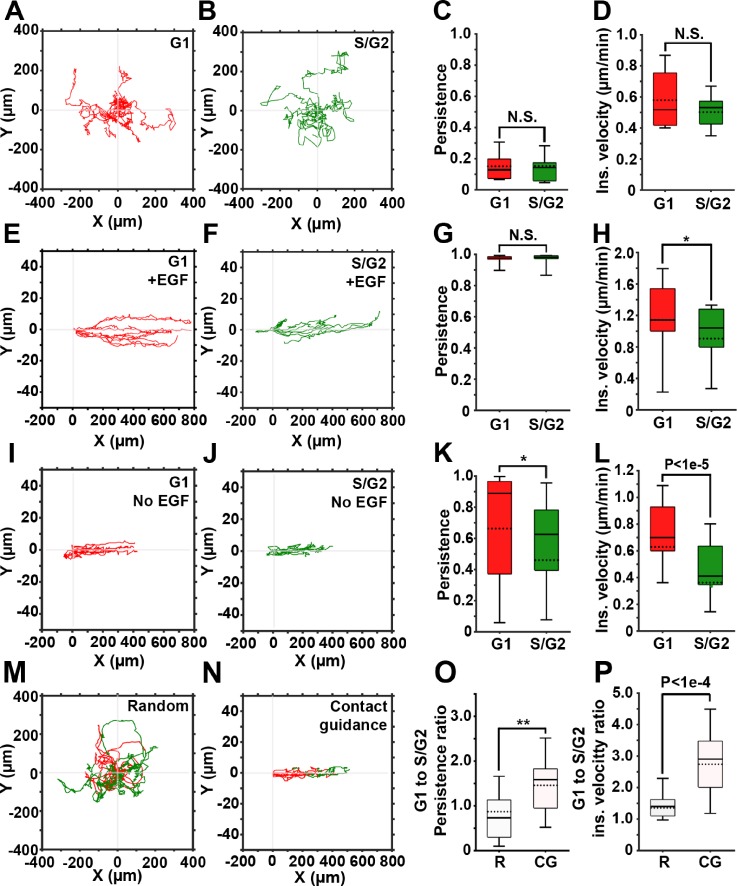
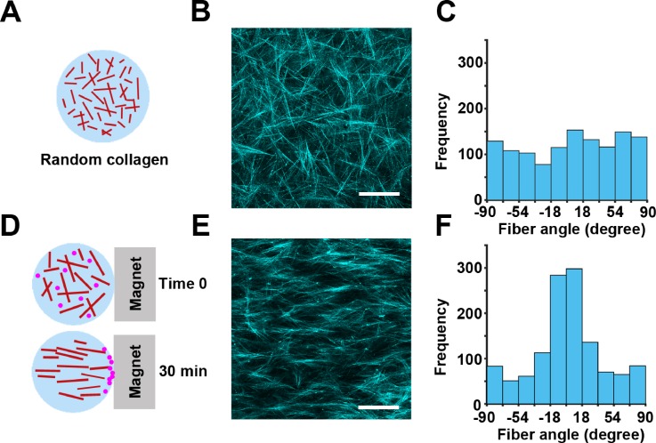
Cancer cell migration is essential for metastasis, during which cancer cells move through the tumor and reach the blood vessels. , cancer cells are exposed to contact guidance and chemotactic cues. Depending on the strength of such cues, cells will migrate in a random or directed manner. While similar cues may also stimulate cell proliferation, it is not clear whether cell cycle progression affects migration of cancer cells and whether this effect is different in random versus directed migration. In this study, we tested the effect of cell cycle progression on contact guided migration in 2D and 3D environments, in the breast carcinoma cell line, FUCCI-MDA-MB-231. The results were quantified from live cell microscopy images using the open source lineage editing and validation image analysis tools (LEVER). In 2D, cells were placed inside 10 m-wide microchannels to stimulate contact guidance, with or without an additional chemotactic gradient of the soluble epidermal growth factor. In 3D, contact guidance was modeled by aligned collagen fibers. In both 2D and 3D, contact guidance was cell cycle-dependent, while the addition of the chemo-attractant gradient in 2D increased cell velocity and persistence in directionally migrating cells, regardless of their cell cycle phases. In both 2D and 3D contact guidance, cells in the G1 phase of the cell cycle outperformed cells in the S/G2 phase in terms of migration persistence and instantaneous velocity. These data suggest that in the presence of contact guidance cues , breast carcinoma cells in the G1 phase of the cell cycle may be more efficient in reaching the neighboring vasculature.
癌细胞迁移是转移的关键,在此过程中癌细胞穿过肿瘤并到达血管。在此期间,癌细胞会受到接触导向和趋化性线索的影响。根据这些线索的强度,细胞将以随机或定向的方式迁移。虽然类似的线索也可能刺激细胞增殖,但尚不清楚细胞周期进程是否会影响癌细胞的迁移,以及这种影响在随机迁移和定向迁移中是否有所不同。在本研究中,我们在乳腺癌细胞系FUCCI-MDA-MB-231中测试了细胞周期进程对二维和三维环境中接触导向迁移的影响。使用开源谱系编辑和验证图像分析工具(LEVER)从活细胞显微镜图像中对结果进行了量化。在二维环境中,将细胞置于10微米宽的微通道内以刺激接触导向,有无可溶性表皮生长因子的额外趋化梯度。在三维环境中,通过排列的胶原纤维模拟接触导向。在二维和三维环境中,接触导向均依赖于细胞周期,而在二维环境中添加趋化剂梯度可增加定向迁移细胞的速度和方向持续性,无论其细胞周期阶段如何。在二维和三维接触导向中,处于细胞周期G1期的细胞在迁移持续性和瞬时速度方面均优于处于S/G2期的细胞。这些数据表明,在存在接触导向线索的情况下,处于细胞周期G1期的乳腺癌细胞可能在到达邻近脉管系统方面更有效。