Nacif Lucas Souto, Paranaguá-Vezozzo Denise Cerqueira, Galvão Flávio Henrique Ferreira, Rocha Manoel S, Andraus Wellington, Carrilho Flair Jose, D'Albuquerque Luiz Carneiro
Liver and Gastrointestinal Transplant Division. Department of Gastroenterology, University of São Paulo School of Medicine, Rua Dr. Enéas de Carvalho Aguiar, 255 -9° andar -sala 9113/9114 CEP 05403-900, São Paulo, Brazil.
BMC Med Imaging. 2015 Sep 18;15:37. doi: 10.1186/s12880-015-0079-7.
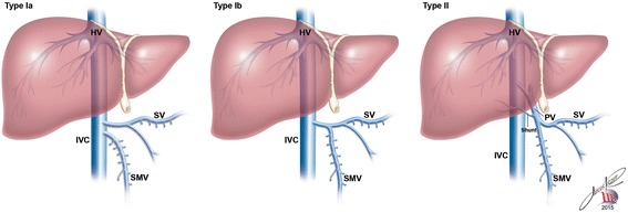
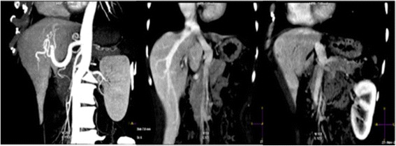
Abernethy malformation is a rare congenital vascular abnormality in which the portal vein bypasses the liver and drains directly into the inferior vena cava. Diagnosis is complex and requires good quality imaging methods to identify details in systemic and portal circulation in order to establish diagnostic confirmation and treatment strategy. In this study we highlight the significance of the use of CT scans and Color Doppler Duplex Ultrasound for the diagnosis, treatment and evolution assessment in two adults with Abernethy malformation.
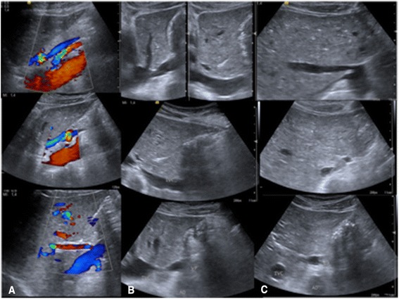
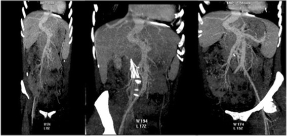
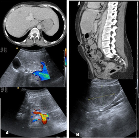
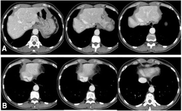
The diagnosis and the treatment of two patients with Abernethy malformation by CT scan and Color Doppler Duplex Ultrasound is described. One patient was submitted to liver transplantation due to chronic liver disease and multiple nodules diagnosed as adenoma. The other patient had normal liver function and a mild neurological and psychomotor dysfunction, therefore we adopted clinical treatment and close liver parenchyma evaluation and nodule surveillance, using an imaging approach involving intercalating CT scan and Color Doppler Duplex Ultrasound every 6 months. We highlight some important direct and indirect findings of non-invasive imaging methods.
Abernethy malformation requires meticulous image diagnosis to improve treatment and avoid iatrogenic procedures. CT scans and Color Doppler Duplex Ultrasound are both efficient methods for diagnosis, treatment planning and evolution assessment of patients with Abernethy malformation.
阿伯内西畸形是一种罕见的先天性血管异常,其中门静脉绕过肝脏,直接引流至下腔静脉。诊断较为复杂,需要高质量的成像方法来识别体循环和门静脉循环中的细节,以确立诊断并制定治疗策略。在本研究中,我们强调了使用CT扫描和彩色多普勒双功能超声对两名患有阿伯内西畸形的成年人进行诊断、治疗及病情进展评估的重要性。
描述了通过CT扫描和彩色多普勒双功能超声对两名阿伯内西畸形患者的诊断和治疗情况。一名患者因慢性肝病及多个诊断为腺瘤的结节而接受了肝移植。另一名患者肝功能正常,有轻度神经和精神运动功能障碍,因此我们采取了临床治疗,并通过每隔6个月进行一次CT扫描和彩色多普勒双功能超声的成像方法,对肝实质进行密切评估并监测结节情况。我们强调了一些非侵入性成像方法的重要直接和间接发现。
阿伯内西畸形需要细致的影像诊断以改善治疗并避免医源性操作。CT扫描和彩色多普勒双功能超声都是诊断、治疗规划及评估阿伯内西畸形患者病情进展的有效方法。