Demirhan Ayşe, Nir Talia M, Zavaliangos-Petropulu Artemis, Jack Clifford R, Weiner Michael W, Bernstein Matt A, Thompson Paul M, Jahanshad Neda
Electronics & Computer Technology, Faculty of Technology, Gazi University, Ankara, Turkey ; Imaging Genetics Center, Keck School of Medicine of USC, Marina del Rey, CA, USA.
Imaging Genetics Center, Keck School of Medicine of USC, Marina del Rey, CA, USA.
Proc IEEE Int Symp Biomed Imaging. 2015 Apr;2015:126-130. doi: 10.1109/ISBI.2015.7163832.
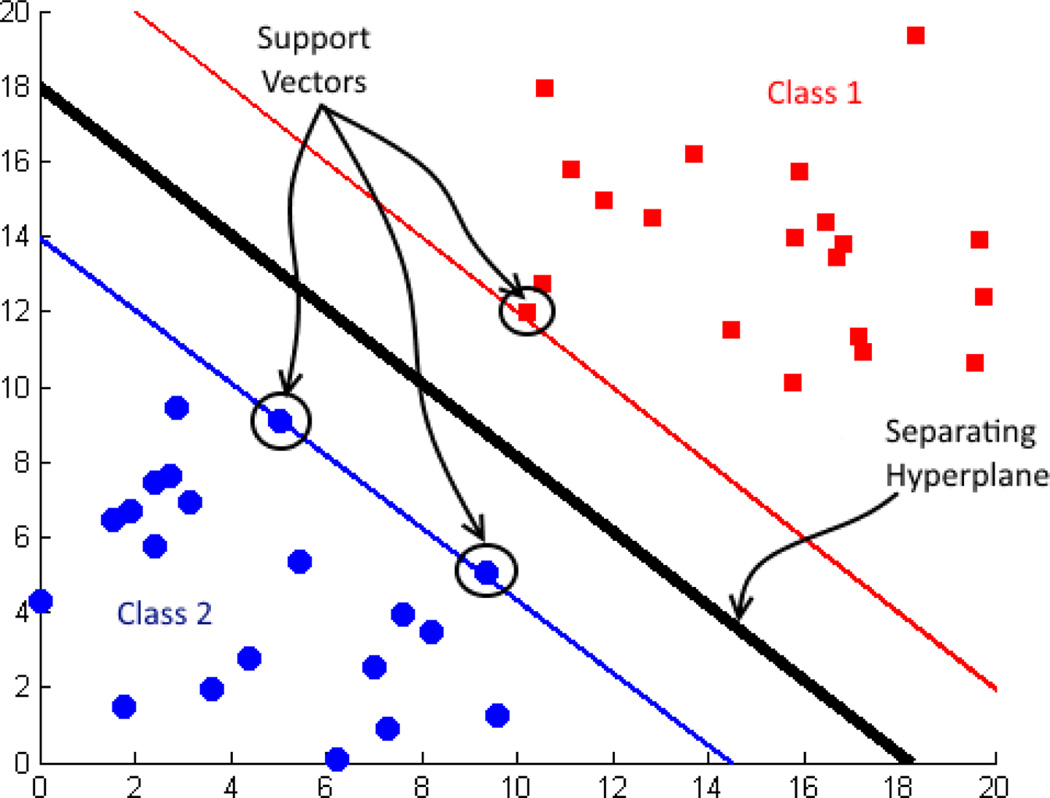
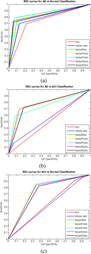
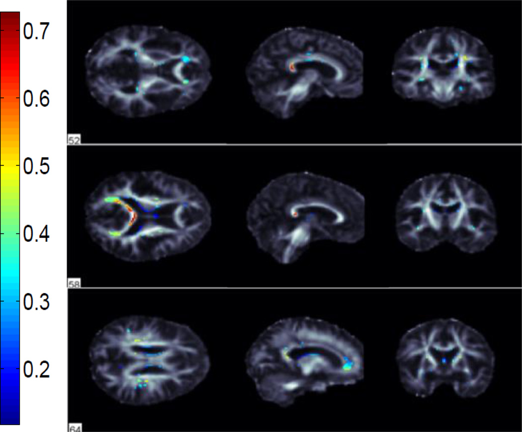
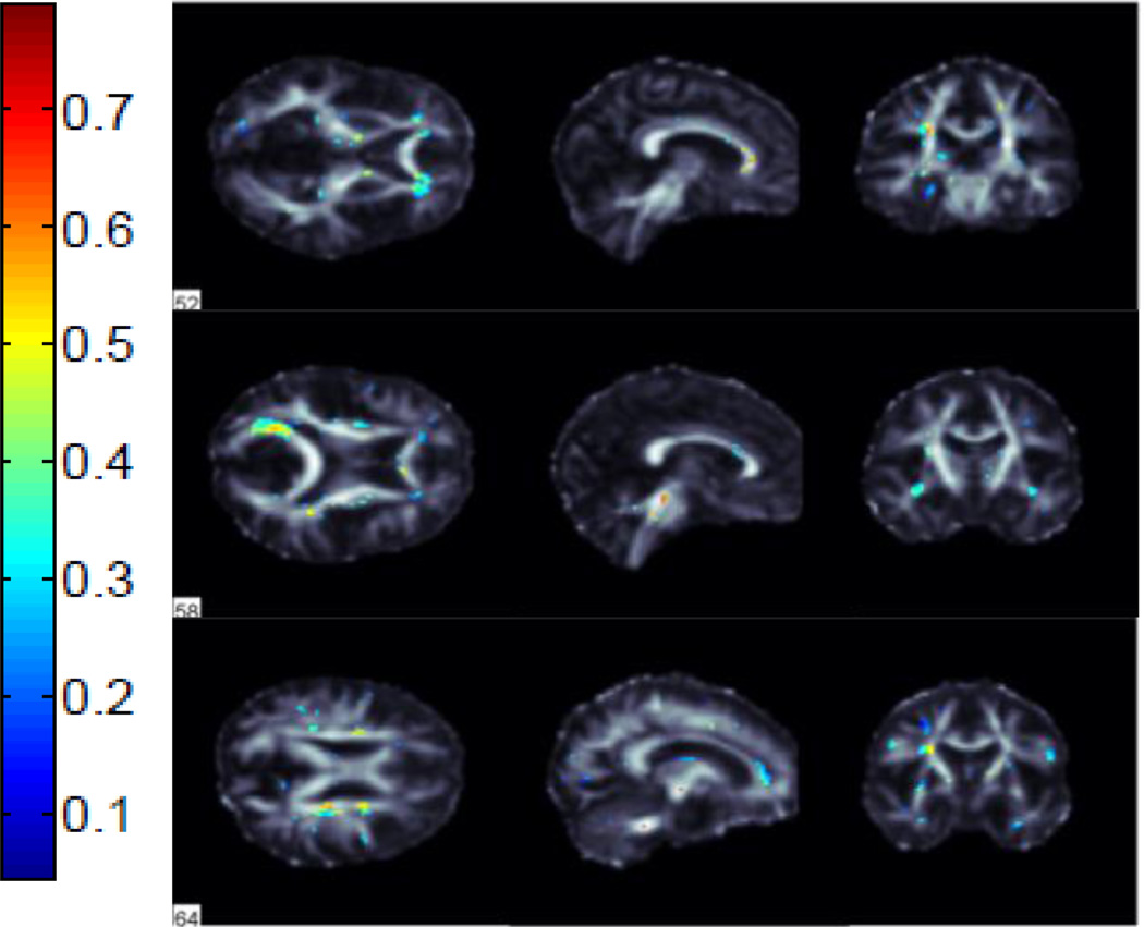
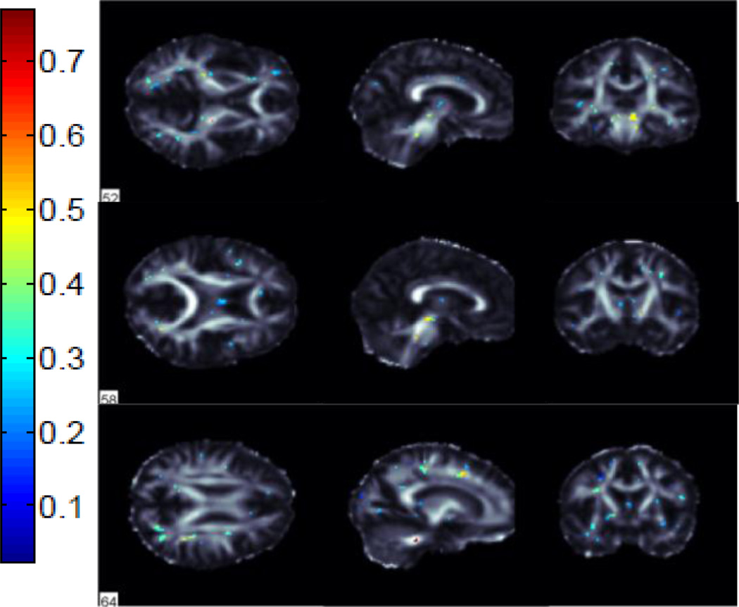
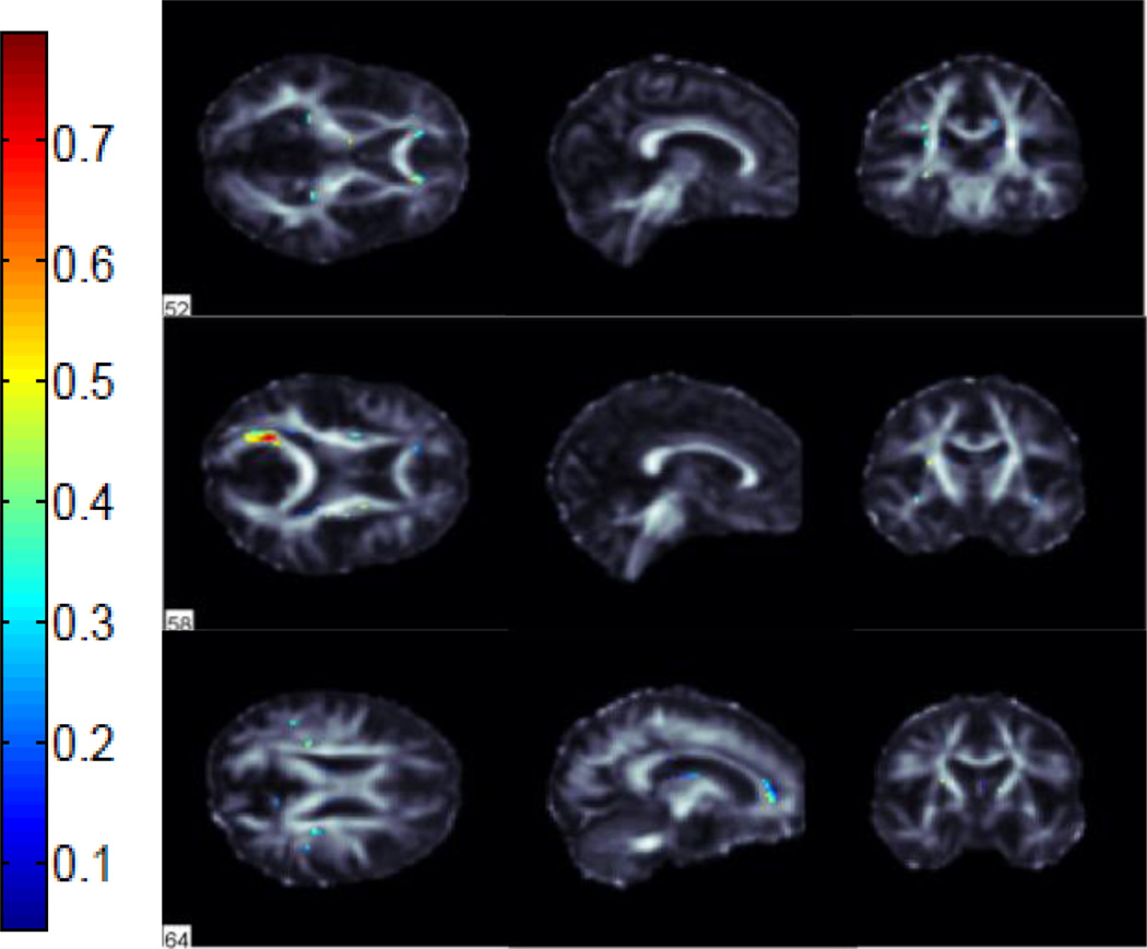
Diffusion tensor imaging (DTI) has recently been added to several large-scale studies of Alzheimer's disease (AD), such as the Alzheimer's Disease Neuroimaging Initiative (ADNI), to investigate white matter (WM) abnormalities not detectable on standard anatomical MRI. Disease effects can be widespread, and the profile of WM abnormalities across tracts is still not fully understood. Here we analyzed image-wide measures from DTI fractional anisotropy (FA) maps to classify AD patients (n=43), mild cognitive impairment (n=114) and cognitively healthy elderly controls (n=70). We used voxelwise maps of FA along with averages in WM regions of interest (ROI) to drive a Support Vector Machine. We further used the ReliefF algorithm to select the most discriminative WM voxels for classification. This improved accuracy for all classification tasks by up to 15%. We found several clusters formed by the ReliefF algorithm, highlighting specific pathways affected in AD but not always captured when analyzing ROIs.
扩散张量成像(DTI)最近已被纳入多项阿尔茨海默病(AD)的大规模研究中,例如阿尔茨海默病神经影像学计划(ADNI),以研究标准解剖磁共振成像(MRI)无法检测到的白质(WM)异常。疾病影响可能很广泛,并且不同脑区白质异常的特征仍未完全了解。在这里,我们分析了DTI分数各向异性(FA)图的全图像测量值,以对AD患者(n = 43)、轻度认知障碍患者(n = 114)和认知健康的老年对照者(n = 70)进行分类。我们使用FA的体素级映射以及感兴趣的白质区域(ROI)的平均值来驱动支持向量机。我们进一步使用ReliefF算法选择最具区分性的白质体素进行分类。这将所有分类任务的准确率提高了多达15%。我们发现了由ReliefF算法形成的几个聚类,突出了AD中受影响的特定通路,但在分析ROI时并不总是能捕捉到。