• 文献检索
  • 文档翻译
  • 深度研究
  • 学术资讯
  • Suppr Zotero 插件Zotero 插件
  • 邀请有礼
  • 套餐&价格
  • 历史记录
应用&插件
Suppr Zotero 插件Zotero 插件浏览器插件Mac 客户端Windows 客户端微信小程序
定价
高级版会员购买积分包购买API积分包
服务
文献检索文档翻译深度研究API 文档MCP 服务
关于我们
关于 Suppr公司介绍联系我们用户协议隐私条款
关注我们

Suppr 超能文献

核心技术专利:CN118964589B侵权必究
粤ICP备2023148730 号-1Suppr @ 2026

文献检索

告别复杂PubMed语法,用中文像聊天一样搜索,搜遍4000万医学文献。AI智能推荐,让科研检索更轻松。

立即免费搜索

文件翻译

保留排版,准确专业,支持PDF/Word/PPT等文件格式,支持 12+语言互译。

免费翻译文档

深度研究

AI帮你快速写综述,25分钟生成高质量综述,智能提取关键信息,辅助科研写作。

立即免费体验

人类颞叶的高场磁共振成像

High-field magnetic resonance imaging of the human temporal lobe.

作者信息

Colon-Perez Luis M, King Michael, Parekh Mansi, Boutzoukas Angelique, Carmona Eduardo, Couret Michelle, Klassen Rosemary, Mareci Thomas H, Carney Paul R

机构信息

Department of Biochemistry and Molecular Biology, University of Florida, Gainesville, FL, USA ; Department of Psychiatry, University of Florida, Gainesville, FL, USA.

Department of Pharmacology and Therapeutics, University of Florida, Gainesville, FL, USA ; Department of Veterans Affairs Medical Center, Gainesville, FL, USA.

出版信息

Neuroimage Clin. 2015 Aug 1;9:58-68. doi: 10.1016/j.nicl.2015.07.005. eCollection 2015.

DOI:10.1016/j.nicl.2015.07.005
PMID:26413472
原文链接:https://pmc.ncbi.nlm.nih.gov/articles/PMC4543219/
Abstract

BACKGROUND

Emerging high-field diffusion weighted MR imaging protocols, along with tractography, can elucidate microstructural changes associated with brain disease at the sub-millimeter image resolution. Epilepsy and other neurological disorders are accompanied by structural changes in the hippocampal formation and associated regions; however, these changes can be subtle and on a much smaller scale than the spatial resolution commonly obtained by current clinical magnetic resonance (MR) protocols in vivo.

METHODS

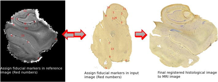
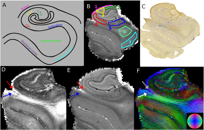
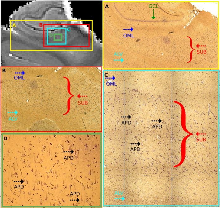
We explored the possibility of studying the organization of fresh tissue with a 17.6 Tesla magnet using diffusion MR imaging and tractography. The mesoscale organization of the temporal lobe was estimated using a fresh unfixed specimen obtained from a subject who underwent anterior temporal lobectomy for medically refractory temporal lobe epilepsy (TLE). Following ex vivo imaging, the tissue was fixed, serial-sectioned, and stained for correlation with imaging.

FINDINGS

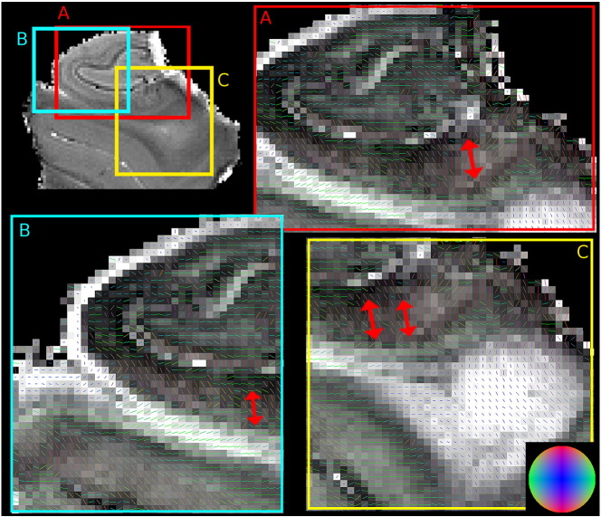
We resolved tissue microstructural organizational features in the temporal lobe from diffusion MR imaging and tractography in fresh tissue.

CONCLUSIONS

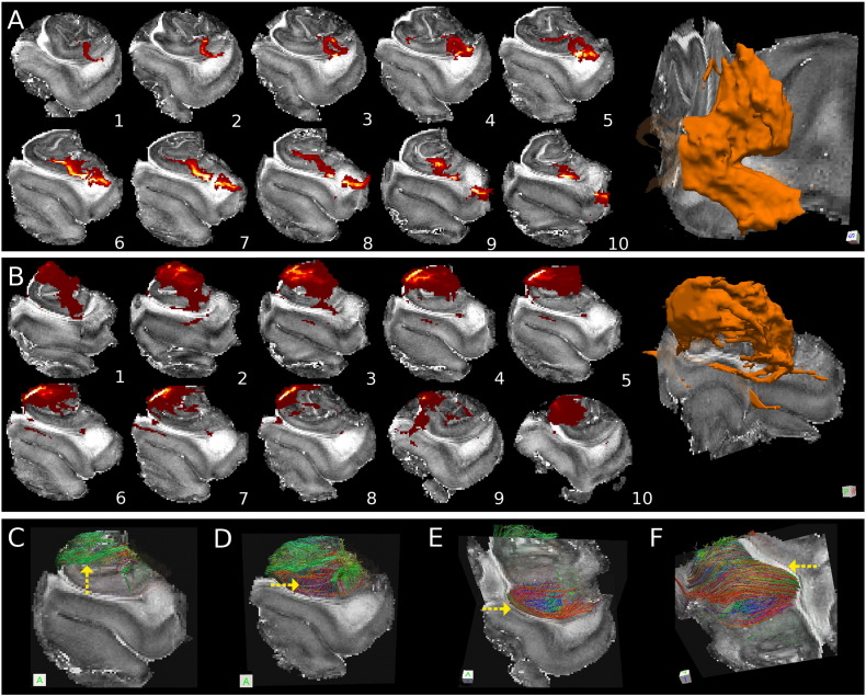
Fresh ex vivo MR imaging, along with tractography, revealed complex intra-temporal structural variation corresponding to neuronal cell body layers, dendritic fields, and axonal projection systems evident histologically. This is the first study to describe in detail the human temporal lobe structural organization using high-field MR imaging and tractography. By preserving the 3-dimensional structures of the hippocampus and surrounding structures, specific changes in anatomy may inform us about the changes that occur in TLE in relation to the disease process and structural underpinnings in epilepsy-related memory dysfunction.

摘要

背景

新兴的高场扩散加权磁共振成像协议,连同纤维束成像,能够在亚毫米图像分辨率下阐明与脑部疾病相关的微观结构变化。癫痫和其他神经系统疾病伴随着海马结构及相关区域的结构变化;然而,这些变化可能很细微,且比当前临床磁共振(MR)协议在体内通常获得的空间分辨率小得多。

方法

我们探讨了使用17.6特斯拉磁体通过扩散磁共振成像和纤维束成像研究新鲜组织结构的可能性。使用从一名因药物难治性颞叶癫痫(TLE)接受前颞叶切除术的受试者获取的新鲜未固定标本,估计颞叶的中尺度结构。在体外成像后,将组织固定、连续切片并染色以与成像结果进行对比。

研究结果

我们通过新鲜组织的扩散磁共振成像和纤维束成像解析了颞叶的组织微观结构特征。

结论

新鲜离体磁共振成像连同纤维束成像揭示了与神经元细胞体层、树突场和轴突投射系统相对应的复杂颞内结构变化,这些在组织学上是明显的。这是第一项使用高场磁共振成像和纤维束成像详细描述人类颞叶结构组织的研究。通过保留海马体及周围结构的三维结构,特定的解剖学变化可能会让我们了解在TLE中与疾病进程及癫痫相关记忆功能障碍的结构基础有关的变化。

https://cdn.ncbi.nlm.nih.gov/pmc/blobs/34df/4543219/4ff0feeac7b3/gr7.jpg
https://cdn.ncbi.nlm.nih.gov/pmc/blobs/34df/4543219/dc8f1347da2e/gr1.jpg
https://cdn.ncbi.nlm.nih.gov/pmc/blobs/34df/4543219/bd3d2bbcd6c3/gr2.jpg
https://cdn.ncbi.nlm.nih.gov/pmc/blobs/34df/4543219/b6acf15ef433/gr3.jpg
https://cdn.ncbi.nlm.nih.gov/pmc/blobs/34df/4543219/729e938db710/gr4.jpg
https://cdn.ncbi.nlm.nih.gov/pmc/blobs/34df/4543219/ec8501279391/gr5.jpg
https://cdn.ncbi.nlm.nih.gov/pmc/blobs/34df/4543219/970e031ca99b/gr6.jpg
https://cdn.ncbi.nlm.nih.gov/pmc/blobs/34df/4543219/4ff0feeac7b3/gr7.jpg

相似文献

1
High-field magnetic resonance imaging of the human temporal lobe.人类颞叶的高场磁共振成像
Neuroimage Clin. 2015 Aug 1;9:58-68. doi: 10.1016/j.nicl.2015.07.005. eCollection 2015.
2
Mesoscale diffusion magnetic resonance imaging of the ex vivo human hippocampus.离体人脑海马体的介观扩散磁共振成像。
Hum Brain Mapp. 2020 Oct 15;41(15):4200-4218. doi: 10.1002/hbm.25119. Epub 2020 Jul 4.
3
Diffusion tensor imaging tractography of Meyer's loop in planning resective surgery for drug-resistant temporal lobe epilepsy.在耐药性颞叶癫痫的手术切除规划中,迈耶袢的扩散张量成像纤维束成像
Epilepsy Res. 2015 Feb;110:95-104. doi: 10.1016/j.eplepsyres.2014.11.020. Epub 2014 Nov 27.
4
Detection of aberrant hippocampal mossy fiber connections: Ex vivo mesoscale diffusion MRI and microtractography with histological validation in a patient with uncontrolled temporal lobe epilepsy.异常海马苔藓纤维连接的检测:一名颞叶癫痫未控制患者的离体中尺度扩散磁共振成像和微观纤维束成像及组织学验证
Hum Brain Mapp. 2016 Feb;37(2):780-95. doi: 10.1002/hbm.23066. Epub 2015 Nov 27.
5
Group-specific regional white matter abnormality revealed in diffusion tensor imaging of medial temporal lobe epilepsy without hippocampal sclerosis.内侧颞叶癫痫伴海马硬化症患者的弥散张量成像显示特定区域的白质异常。
Epilepsia. 2010 Apr;51(4):529-35. doi: 10.1111/j.1528-1167.2009.02327.x. Epub 2009 Oct 8.
6
Connectivity and tissue microstructural alterations in right and left temporal lobe epilepsy revealed by diffusion spectrum imaging.扩散光谱成像揭示的左右颞叶癫痫中的连通性和组织微观结构改变
Neuroimage Clin. 2014 Aug 1;5:349-58. doi: 10.1016/j.nicl.2014.07.013. eCollection 2014.
7
7T MRI features in control human hippocampus and hippocampal sclerosis: an ex vivo study with histologic correlations.7T MRI 特征在正常人类海马体和海马硬化症中的表现:一项具有组织学相关性的离体研究。
Epilepsia. 2014 Dec;55(12):2003-16. doi: 10.1111/epi.12828. Epub 2014 Nov 3.
8
Diffusion tensor imaging tractography parameters of limbic system bundles in temporal lobe epilepsy patients.颞叶癫痫患者边缘系统束的弥散张量成像轨迹参数。
J Magn Reson Imaging. 2012 Sep;36(3):561-8. doi: 10.1002/jmri.23678. Epub 2012 May 2.
9
Functional MRI, resting state fMRI, and DTI for predicting verbal fluency outcome following resective surgery for temporal lobe epilepsy.功能磁共振成像、静息态功能磁共振成像和弥散张量成像用于预测颞叶癫痫切除术后的言语流畅性结果。
J Neurosurg. 2016 Apr;124(4):929-37. doi: 10.3171/2014.9.JNS131422. Epub 2015 Sep 25.
10
In vivo mapping of macroscopic neuronal projections in the mouse hippocampus using high-resolution diffusion MRI.使用高分辨率扩散磁共振成像对小鼠海马体中的宏观神经元投射进行体内图谱绘制。
Neuroimage. 2016 Jan 15;125:84-93. doi: 10.1016/j.neuroimage.2015.10.051. Epub 2015 Oct 21.

引用本文的文献

1
Reduced structural connectivity of the medial temporal lobe including the perforant path is associated with aging and verbal memory impairment.内侧颞叶(包括穿通纤维束)的结构连接减少与衰老和言语记忆损伤有关。
Neurobiol Aging. 2023 Jan;121:119-128. doi: 10.1016/j.neurobiolaging.2022.10.012. Epub 2022 Nov 9.
2
Hippocampal dentate gyrus integrity revealed with ultrahigh resolution diffusion imaging predicts memory performance in older adults.超高分辨率弥散成像显示海马齿状回完整可预测老年人的记忆表现。
Hippocampus. 2022 Sep;32(9):627-638. doi: 10.1002/hipo.23456. Epub 2022 Jul 15.
3
Mesoscale diffusion magnetic resonance imaging of the ex vivo human hippocampus.

本文引用的文献

1
7T MRI features in control human hippocampus and hippocampal sclerosis: an ex vivo study with histologic correlations.7T MRI 特征在正常人类海马体和海马硬化症中的表现:一项具有组织学相关性的离体研究。
Epilepsia. 2014 Dec;55(12):2003-16. doi: 10.1111/epi.12828. Epub 2014 Nov 3.
2
Stroke and the connectome: how connectivity guides therapeutic intervention.中风与连接组学:连接如何指导治疗干预。
Neuron. 2014 Sep 17;83(6):1354-68. doi: 10.1016/j.neuron.2014.08.052.
3
Engineering the next generation of clinical deep brain stimulation technology.
离体人脑海马体的介观扩散磁共振成像。
Hum Brain Mapp. 2020 Oct 15;41(15):4200-4218. doi: 10.1002/hbm.25119. Epub 2020 Jul 4.
4
Quantitative synchrotron X-ray tomography of the material-tissue interface in rat cortex implanted with neural probes.大鼠皮层植入神经探针后的材料-组织界面的定量同步辐射 X 射线断层扫描。
Sci Rep. 2019 May 21;9(1):7646. doi: 10.1038/s41598-019-42544-9.
5
Cognition and connectomes in nondementia idiopathic Parkinson's disease.非痴呆型特发性帕金森病的认知与脑连接组
Netw Neurosci. 2018 Mar 1;2(1):106-124. doi: 10.1162/NETN_a_00027. eCollection 2018 Spring.
6
Post-mortem inference of the human hippocampal connectivity and microstructure using ultra-high field diffusion MRI at 11.7 T.利用 11.7T 超高场弥散 MRI 对人体海马体连接和微观结构进行死后推断。
Brain Struct Funct. 2018 Jun;223(5):2157-2179. doi: 10.1007/s00429-018-1617-1. Epub 2018 Jan 31.
7
The hippocampus: detailed assessment of normative two-dimensional measurements, signal intensity, and subfield conspicuity on routine 3T T2-weighted sequences.海马体:在常规3T T2加权序列上对标准化二维测量、信号强度及亚区域清晰度的详细评估
Surg Radiol Anat. 2017 Oct;39(10):1149-1159. doi: 10.1007/s00276-017-1843-x. Epub 2017 Mar 12.
8
Toward 20 T magnetic resonance for human brain studies: opportunities for discovery and neuroscience rationale.迈向用于人类大脑研究的20特斯拉磁共振成像:发现机遇与神经科学原理
MAGMA. 2016 Jun;29(3):617-39. doi: 10.1007/s10334-016-0561-4. Epub 2016 May 18.
9
Robust diffusion tensor imaging by spatiotemporal encoding: Principles and in vivo demonstrations.通过时空编码实现的稳健扩散张量成像:原理与体内演示
Magn Reson Med. 2017 Mar;77(3):1124-1133. doi: 10.1002/mrm.26197. Epub 2016 Mar 10.
工程下一代临床深部脑刺激技术。
Brain Stimul. 2015 Jan-Feb;8(1):21-6. doi: 10.1016/j.brs.2014.07.039. Epub 2014 Jul 30.
4
Interlamellar CA1 network in the hippocampus.海马体中的层间CA1网络。
Proc Natl Acad Sci U S A. 2014 Sep 2;111(35):12919-24. doi: 10.1073/pnas.1405468111. Epub 2014 Aug 19.
5
Evaluating contextual processing in diffusion MRI: application to optic radiation reconstruction for epilepsy surgery.评估扩散磁共振成像中的上下文处理:在癫痫手术视辐射重建中的应用。
PLoS One. 2014 Jul 31;9(7):e101524. doi: 10.1371/journal.pone.0101524. eCollection 2014.
6
A mesoscale connectome of the mouse brain.小鼠大脑的介观连接组图谱
Nature. 2014 Apr 10;508(7495):207-14. doi: 10.1038/nature13186. Epub 2014 Apr 2.
7
Disrupted anatomic white matter network in left mesial temporal lobe epilepsy.左侧颞叶内侧癫痫的解剖结构白质网络紊乱。
Epilepsia. 2014 May;55(5):674-682. doi: 10.1111/epi.12581. Epub 2014 Mar 20.
8
Early detection of widespread progressive brain injury after cardiac arrest: a single case DTI and post-mortem histology study.心脏骤停后广泛性进行性脑损伤的早期检测:单病例DTI及尸检组织学研究
PLoS One. 2014 Mar 14;9(3):e92103. doi: 10.1371/journal.pone.0092103. eCollection 2014.
9
Quantitative mapping of trimethyltin injury in the rat brain using magnetic resonance histology.使用磁共振组织学对大鼠脑内三甲基锡损伤进行定量映射。
Neurotoxicology. 2014 May;42:12-23. doi: 10.1016/j.neuro.2014.02.009. Epub 2014 Mar 11.
10
Quantifying diffusion MRI tractography of the corticospinal tract in brain tumors with deterministic and probabilistic methods.应用确定性和概率性方法对脑肿瘤皮质脊髓束的弥散 MRI 轨迹进行量化。
Neuroimage Clin. 2013 Aug 20;3:361-8. doi: 10.1016/j.nicl.2013.08.008. eCollection 2013.