Gerdes Jan S, Walther Ernst U, Jaganjac Suad, Makrigeorgi-Butera Maria, Meuth Sven G, Deppe Michael
Department of Neurology, Schön Klinik Hamburg Eilbek, Hamburg, Germany.
Department of Radiology, Schön Klinik Hamburg Eilbek, Hamburg, Germany.
PLoS One. 2014 Mar 14;9(3):e92103. doi: 10.1371/journal.pone.0092103. eCollection 2014.
We tested the hypothesis in sense of a proof of principle that white matter (WM) degeneration after cardiopulmonary arrest (CPA) can be assessed much earlier by diffusion tensor imaging (DTI) than by conventional MRI.
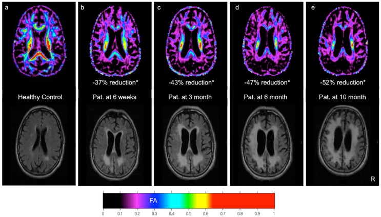
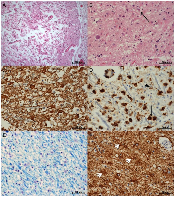
We performed DTI and T2-weighted FLAIR imaging over four serial acquisitions of a 76-year-old man with unresponsive wakefulness syndrome at day 41, 75, 173 and 284 after CPA. DTI was also performed in ten healthy control subjects. Fractional anisotropy (FA) derived from DTI was assessed in eleven regions of interest within the cerebral white matter (WM) and compared with post-mortem neuropathological findings.
In contrast to conventional FLAIR images that revealed only circumscribed WM damage, the first DTI demonstrated significant reduction of FA across the whole WM. The following FLAIR images (MRI 2-4) revealed increasing atrophy and leukoaraiosis paralleled by clinical deterioration with reduction of wakefulness and intractable seizures. Neuropathological findings confirmed the widespread and marked brain injury following CPA.
DTI may help to evaluate microstructural brain damage following CPA and may have predictive value for further evolution of cerebral degeneration in the chronic phase after CPA.
我们从原理验证的角度检验了这一假设,即与传统磁共振成像(MRI)相比,弥散张量成像(DTI)能够更早地评估心脏骤停(CPA)后的白质(WM)退变情况。
我们对一名76岁无反应觉醒综合征男性患者在CPA后第41天、75天、173天和284天进行了4次连续的DTI和T2加权液体衰减反转恢复(FLAIR)成像检查。还对10名健康对照者进行了DTI检查。对脑白质(WM)内11个感兴趣区域的DTI衍生分数各向异性(FA)进行评估,并与死后神经病理学结果进行比较。
与仅显示局限性WM损伤的传统FLAIR图像不同,首次DTI显示整个WM的FA显著降低。随后的FLAIR图像(MRI 2 - 4)显示萎缩和脑白质疏松增加,同时临床病情恶化,觉醒减少和顽固性癫痫发作。神经病理学结果证实了CPA后广泛而明显的脑损伤。
DTI可能有助于评估CPA后脑微观结构损伤,并可能对CPA后慢性期脑退变的进一步发展具有预测价值。