Department of Dermatology, Tokyo Medical University 6-7-1 Nishi-Shinjuku, Shinjuku-ku, Tokyo 160-0023, Japan.
Atopy (Allergy) Research Center, Juntendo University School of Medicine 2-1-1 Hongo, Bunkyo-ku, Tokyo 113-8421, Japan.
Immun Inflamm Dis. 2015 Sep;3(3):196-208. doi: 10.1002/iid3.59. Epub 2015 May 6.
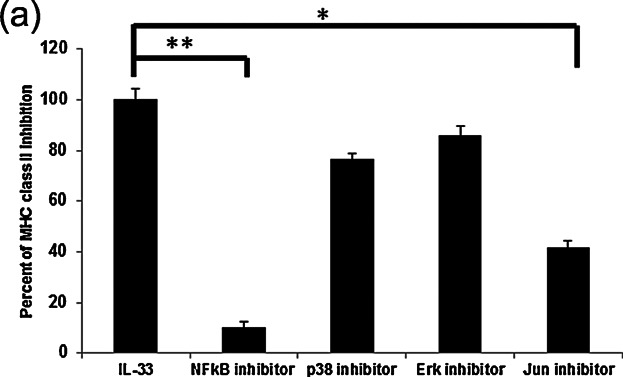
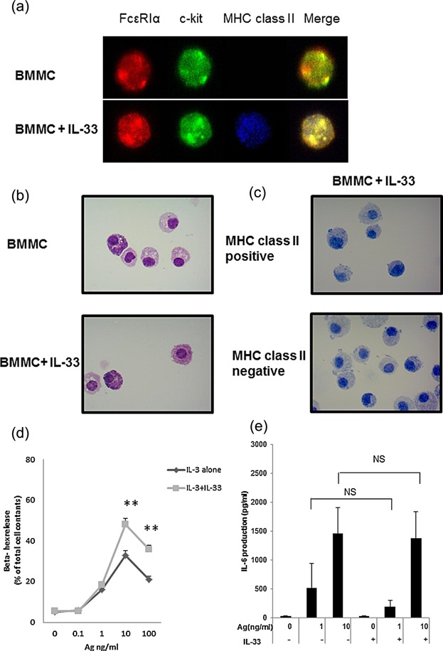
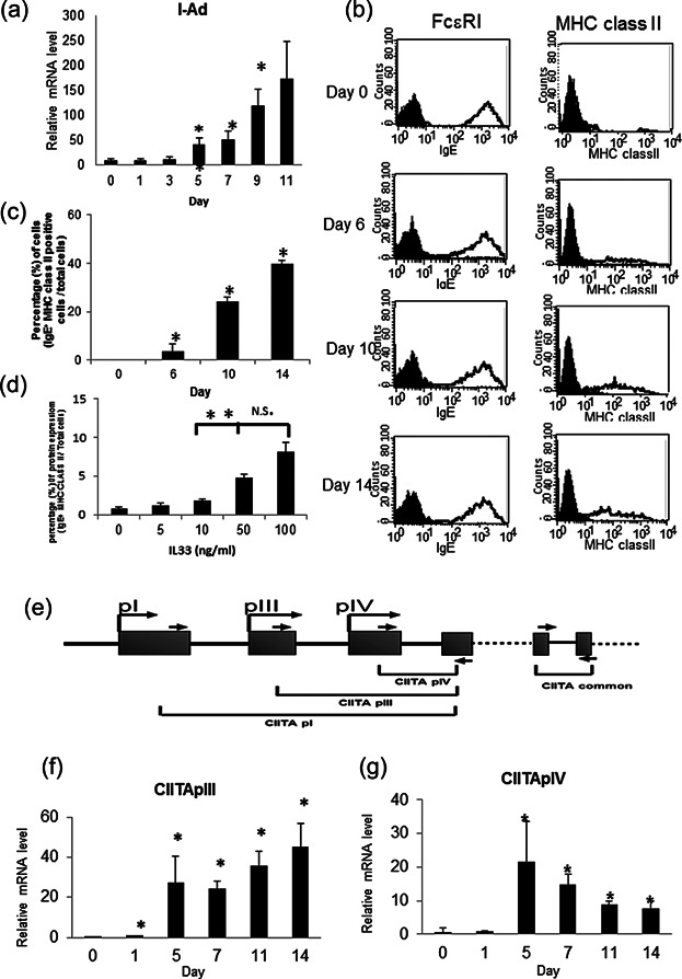
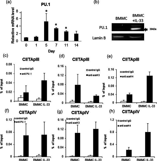
Mast cells (MCs), recognized as tissue-resident cells of hematopoietic origin, are involved in cellular and pathological manifestations of allergic disorders including atopic dermatitis. IL-33, a member of the IL-1 cytokine family, activates Th2-type immune responses, and promotes the degranulation and maturation of MCs. However, it is uncertain whether IL-33 treatment induces mature mast cells to acquire the characteristics of the monocyte-dendritic cell lineage.We investigated the effect of IL-33 on the MHC class II expression and function of murine mast cells. IL-33-treated mature murine bone marrow-derived mast cells (BMMCs) were analyzed by FACS, real-time PCR, chromatin immunoprecipitation (ChIP) assay, and Western blotting. The morphology and degranulation activity of BMMCs and T-cell activation by BMMCs were also examined. BMMCs treated with IL-33 for 10 days induced cell surface expression of the MHC class II protein, whereas the expression of FcεRI and c-kit was not affected by IL-33. The expression of CIITA, driven from pIII and pIV, was up-regulated in IL-33-treated BMMCs. The amount of PU.1 mRNA and protein significantly increased in IL-33-treated BMMCs. The ChIP assay showed PU.1 binding to CIITA pIII, and enhanced histone acetylation due to IL-33 treatment. Syngeneic T cells were activated by co-culture with IL-33-treated BMMCs, although the expression of the co-stimulatory molecules, CD40, CD80, CD86, and PDL-1, was not detected. Mast cells express MHC class II after prolonged exposure to IL-33, probably due to enhanced recruitment of PU.1 to CIITA pIII, resulting in transactivation of CIITA and MHC class II. IL-33 is an important cytokine in allergic disorders. Mast cells have the ability to express MHC class II after prolonged exposure to IL-33 in a murine model. IL-33 holds a key to understanding the etiology of atopic dermatitis.
肥大细胞(MCs)作为造血来源的组织驻留细胞,参与包括特应性皮炎在内的过敏疾病的细胞和病理表现。IL-33 是 IL-1 细胞因子家族的成员,可激活 Th2 型免疫反应,并促进 MCs 的脱颗粒和成熟。然而,IL-33 治疗是否诱导成熟肥大细胞获得单核细胞-树突状细胞谱系的特征尚不确定。我们研究了 IL-33 对鼠类肥大细胞 MHC Ⅱ类表达和功能的影响。通过 FACS、实时 PCR、染色质免疫沉淀(ChIP)分析和 Western blot 分析 IL-33 处理的成熟鼠骨髓来源肥大细胞(BMMCs)。还检查了 BMMC 的形态和脱颗粒活性以及 BMMC 对 T 细胞的激活作用。用 IL-33 处理 10 天的 BMMC 诱导 MHC Ⅱ类蛋白的细胞表面表达,而 IL-33 不影响 FcεRI 和 c-kit 的表达。从 pIII 和 pIV 驱动的 CIITA 的表达在 IL-33 处理的 BMMC 中上调。IL-33 处理的 BMMC 中 PU.1 mRNA 和蛋白的量显著增加。ChIP 分析显示 PU.1 与 CIITA pIII 结合,并因 IL-33 处理而增强组蛋白乙酰化。与 IL-33 处理的 BMMC 共培养可激活同种型 T 细胞,尽管未检测到共刺激分子 CD40、CD80、CD86 和 PDL-1 的表达。肥大细胞在长时间暴露于 IL-33 后表达 MHC Ⅱ类,可能是由于 PU.1 向 CIITA pIII 的募集增强,导致 CIITA 和 MHC Ⅱ类的反式激活。IL-33 是过敏疾病中的重要细胞因子。在鼠模型中,肥大细胞在长时间暴露于 IL-33 后具有表达 MHC Ⅱ类的能力。IL-33 是理解特应性皮炎病因的关键。