• 文献检索
  • 文档翻译
  • 深度研究
  • 学术资讯
  • Suppr Zotero 插件Zotero 插件
  • 邀请有礼
  • 套餐&价格
  • 历史记录
应用&插件
Suppr Zotero 插件Zotero 插件浏览器插件Mac 客户端Windows 客户端微信小程序
定价
高级版会员购买积分包购买API积分包
服务
文献检索文档翻译深度研究API 文档MCP 服务
关于我们
关于 Suppr公司介绍联系我们用户协议隐私条款
关注我们

Suppr 超能文献

核心技术专利:CN118964589B侵权必究
粤ICP备2023148730 号-1Suppr @ 2026

文献检索

告别复杂PubMed语法,用中文像聊天一样搜索,搜遍4000万医学文献。AI智能推荐,让科研检索更轻松。

立即免费搜索

文件翻译

保留排版,准确专业,支持PDF/Word/PPT等文件格式,支持 12+语言互译。

免费翻译文档

深度研究

AI帮你快速写综述,25分钟生成高质量综述,智能提取关键信息,辅助科研写作。

立即免费体验

皮质-皮质连接模式揭示了狨猴中与背侧V2相邻的两个不同的视觉皮质区域。

Corticocortical connection patterns reveal two distinct visual cortical areas bordering dorsal V2 in marmoset monkey.

作者信息

Jeffs Janelle, Federer Frederick, Angelucci Alessandra

机构信息

Department of Ophthalmology and Visual Science,Moran Eye Institute,University of Utah,Salt Lake City,Utah 84132.

出版信息

Vis Neurosci. 2015 Jan;32:E012. doi: 10.1017/S0952523815000097.

DOI:10.1017/S0952523815000097
PMID:26423121
原文链接:https://pmc.ncbi.nlm.nih.gov/articles/PMC5301919/
Abstract

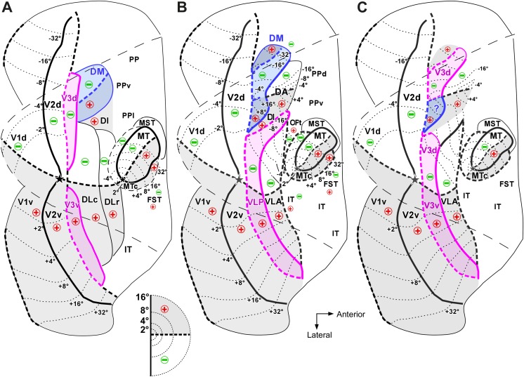
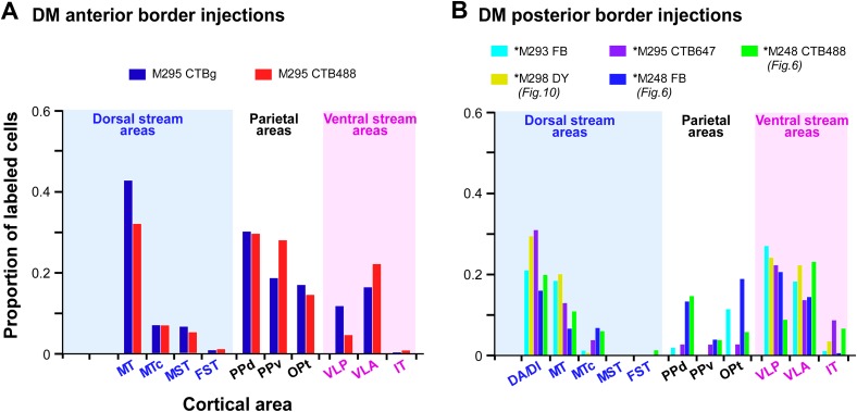
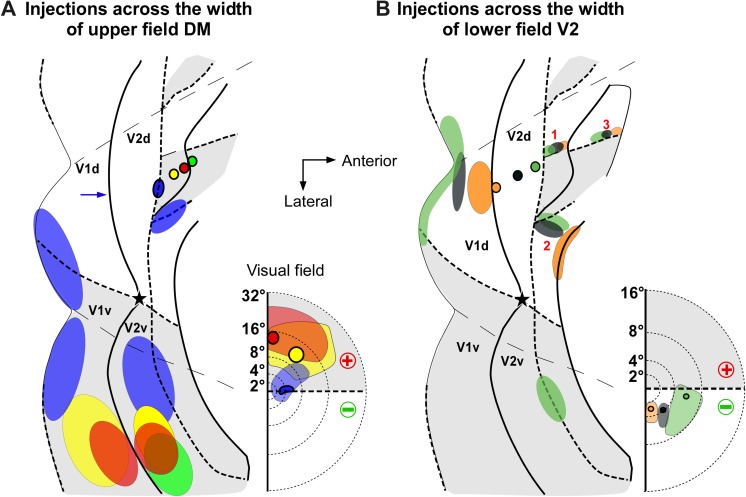
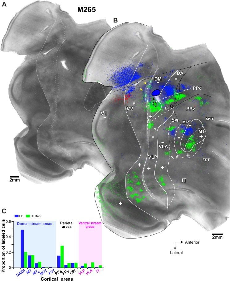
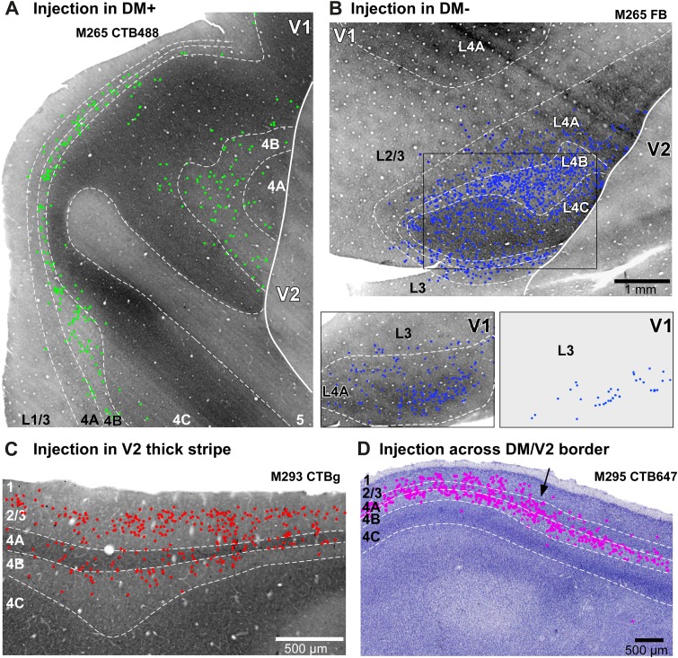
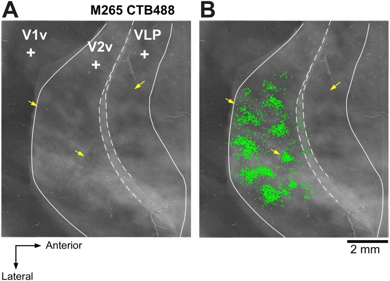
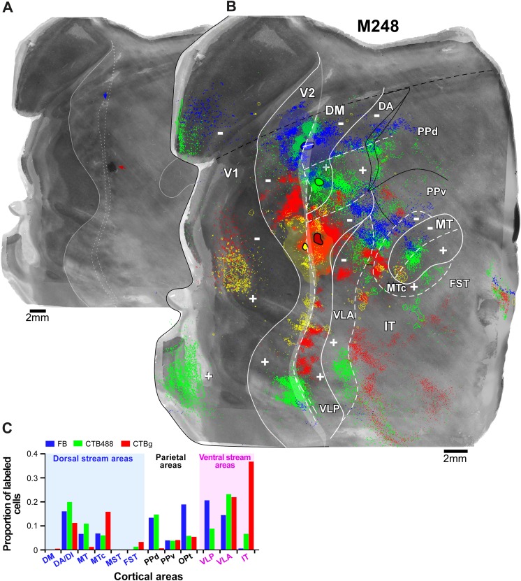
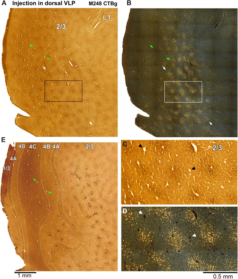
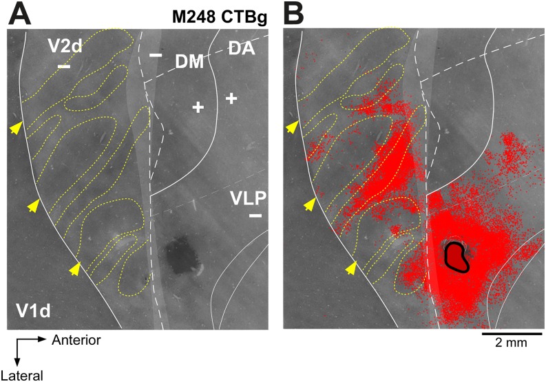
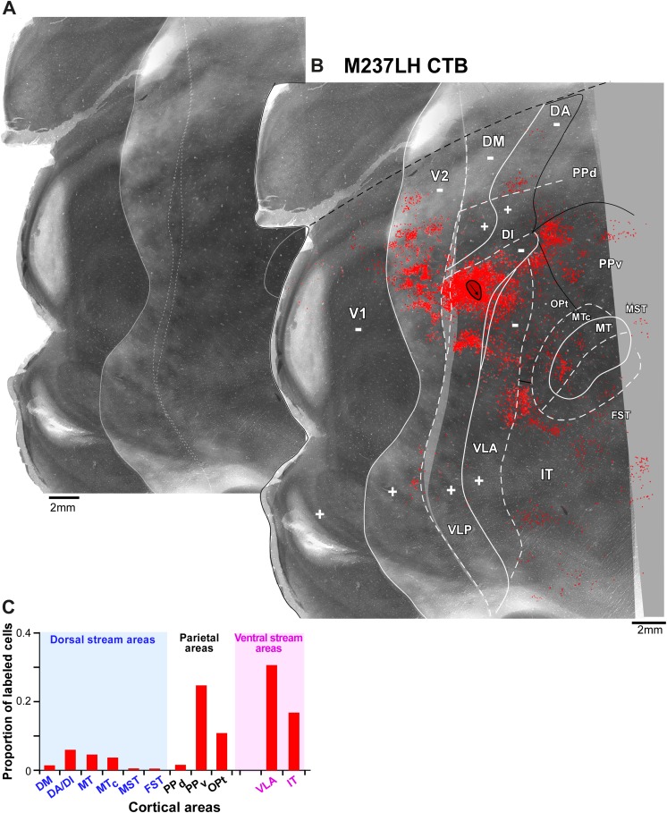
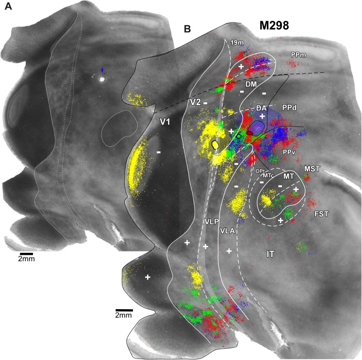
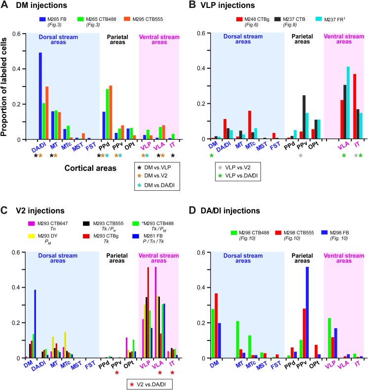
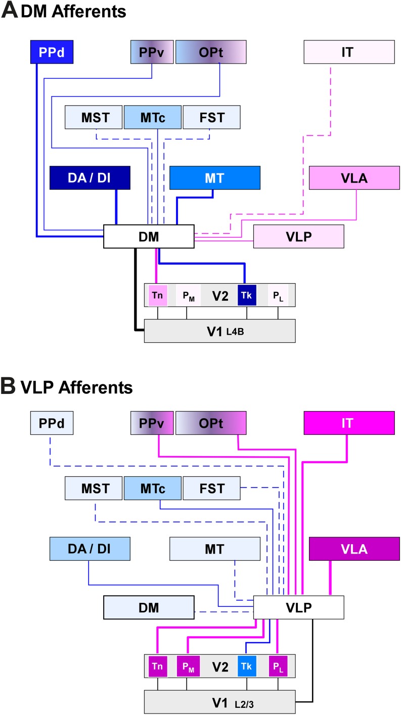
The organization of the cortex located immediately anterior to the second visual area (V2), i.e., the third tier visual cortex, remains controversial, especially in New World primates. In particular, there is lack of consensus regarding the exact location and extent of the lower visual quadrant representation of the third visual area V3 (or ventrolateral posterior -VLP - of a different nomenclature). Microelectrode and connectional mapping studies have revealed the existence of an upper visual quadrant representation abutting dorsal V2 anteriorly, and bordered medially and laterally by representations of the lower visual quadrant. It remains unclear whether these lower field regions are both part of a single area V3, which is split into two patches by an interposed region of upper field representation, or whether they are the lower field representations of two different areas, the dorsomedial area (DM) and area V3/VLP, respectively. To address this question, we quantitatively analyzed the patterns of corticocortical afferent connections labeled by tracer injections targeted to these two lower field regions in the dorsal aspect of the third tier cortex. We found different inter-areal connectivity patterns arising from these two regions, strongly suggesting that they belong to two different visual areas. In particular, our results indicate that the dorsal aspect of the third tier cortex consists of two distinct areas: a full area DM, representing the lower quadrant medially, and the upper quadrant laterally, and the lower quadrant representation of V3/VLP, located laterally to upper field DM. DM is predominantly connected with areas of the dorsal visual stream, and V3/VLP with areas of the ventral stream. These results prompt further functional investigations of the third tier cortex, as previous studies of this cortical territory may have pooled response properties of two very different areas into a single area V3.

摘要

紧邻第二视觉区(V2)前方的皮质组织,即第三层视觉皮质,其组织结构仍存在争议,在新大陆灵长类动物中尤为如此。特别是,关于第三视觉区V3(或不同命名法中的腹外侧后区-VLP-)在下视觉象限的精确位置和范围,目前尚无共识。微电极和连接图谱研究揭示了在背侧V2前方存在一个上视觉象限表征,其内侧和外侧由下视觉象限表征界定。目前尚不清楚这些下视野区域是属于单个区域V3的一部分,该区域被上视野表征的插入区域分成两个斑块,还是它们分别是两个不同区域,即背内侧区(DM)和V3/VLP区的下视野表征。为了解决这个问题,我们定量分析了通过向第三层皮质背侧的这两个下视野区域注射示踪剂标记的皮质皮质传入连接模式。我们发现这两个区域产生了不同的区域间连接模式,强烈表明它们属于两个不同的视觉区域。特别是,我们的结果表明,第三层皮质的背侧由两个不同的区域组成:一个完整的DM区域,在内侧代表下象限,在外侧代表上象限,以及V3/VLP的下象限表征,位于上视野DM的外侧。DM主要与背侧视觉流区域相连,而V3/VLP与腹侧流区域相连。这些结果促使对第三层皮质进行进一步的功能研究,因为此前对该皮质区域的研究可能将两个非常不同区域的反应特性汇总到了单个区域V3中。

https://cdn.ncbi.nlm.nih.gov/pmc/blobs/36e2/5678272/380342c90798/S0952523815000097_fig13.jpg
https://cdn.ncbi.nlm.nih.gov/pmc/blobs/36e2/5678272/dd22f0f3fb44/S0952523815000097_fig1.jpg
https://cdn.ncbi.nlm.nih.gov/pmc/blobs/36e2/5678272/4c1de1c7088a/S0952523815000097_fig2.jpg
https://cdn.ncbi.nlm.nih.gov/pmc/blobs/36e2/5678272/d2fc00bdfe7e/S0952523815000097_fig3.jpg
https://cdn.ncbi.nlm.nih.gov/pmc/blobs/36e2/5678272/f6d63f188547/S0952523815000097_fig4.jpg
https://cdn.ncbi.nlm.nih.gov/pmc/blobs/36e2/5678272/f5b21540ebf7/S0952523815000097_fig5.jpg
https://cdn.ncbi.nlm.nih.gov/pmc/blobs/36e2/5678272/d9092fdf1bea/S0952523815000097_fig6.jpg
https://cdn.ncbi.nlm.nih.gov/pmc/blobs/36e2/5678272/f6c22addc1c4/S0952523815000097_fig7.jpg
https://cdn.ncbi.nlm.nih.gov/pmc/blobs/36e2/5678272/add85815e2c9/S0952523815000097_fig8.jpg
https://cdn.ncbi.nlm.nih.gov/pmc/blobs/36e2/5678272/bc2670cfc33f/S0952523815000097_fig9.jpg
https://cdn.ncbi.nlm.nih.gov/pmc/blobs/36e2/5678272/441631762de4/S0952523815000097_fig10.jpg
https://cdn.ncbi.nlm.nih.gov/pmc/blobs/36e2/5678272/6863437b8940/S0952523815000097_fig11.jpg
https://cdn.ncbi.nlm.nih.gov/pmc/blobs/36e2/5678272/bf596d37df6e/S0952523815000097_fig12.jpg
https://cdn.ncbi.nlm.nih.gov/pmc/blobs/36e2/5678272/380342c90798/S0952523815000097_fig13.jpg

相似文献

1
Corticocortical connection patterns reveal two distinct visual cortical areas bordering dorsal V2 in marmoset monkey.皮质-皮质连接模式揭示了狨猴中与背侧V2相邻的两个不同的视觉皮质区域。
Vis Neurosci. 2015 Jan;32:E012. doi: 10.1017/S0952523815000097.
2
High-resolution mapping of anatomical connections in marmoset extrastriate cortex reveals a complete representation of the visual field bordering dorsal V2.高分辨率映射狨猴外纹状皮层的解剖连接,揭示了紧邻 V2 背侧的视野的完整代表。
Cereb Cortex. 2013 May;23(5):1126-47. doi: 10.1093/cercor/bhs088. Epub 2012 Apr 20.
3
Resolving the organization of the third tier visual cortex in primates: a hypothesis-based approach.解析灵长类动物第三层视觉皮层的组织结构:一种基于假设的方法。
Vis Neurosci. 2015 Jan;32:E010. doi: 10.1017/S0952523815000073.
4
Resolving the organization of the territory of the third visual area: a new proposal.解析第三视觉区域的领土组织:一项新提议。
Vis Neurosci. 2015 Jan;32:E016. doi: 10.1017/S0952523815000152.
5
Resolving the organization of the New World monkey third visual complex: the dorsal extrastriate cortex of the marmoset (Callithrix jacchus).解析新大陆猴第三视觉复合体的组织结构:狨猴(绢毛猴)的背侧纹外皮层。
J Comp Neurol. 2005 Mar 7;483(2):164-91. doi: 10.1002/cne.20412.
6
Topographic Organization of the 'Third-Tier' Dorsomedial Visual Cortex in the Macaque.猴大脑第三级背侧视皮质的拓扑组织。
J Neurosci. 2019 Jul 3;39(27):5311-5325. doi: 10.1523/JNEUROSCI.0085-19.2019. Epub 2019 Apr 29.
7
Visual areas in lateral and ventral extrastriate cortices of the marmoset monkey.狨猴外侧和腹侧纹外皮层的视觉区域。
J Comp Neurol. 2000 Jul 10;422(4):621-51. doi: 10.1002/1096-9861(20000710)422:4<621::aid-cne10>3.0.co;2-e.
8
Connectional and architectonic evidence for dorsal and ventral V3, and dorsomedial area in marmoset monkeys.狨猴背侧和腹侧V3以及背内侧区域的连接和构筑学证据。
J Neurosci. 2001 Jan 1;21(1):249-61. doi: 10.1523/JNEUROSCI.21-01-00249.2001.
9
Visual field representation in striate and prestriate cortices of a prosimian primate (Galago garnetti).一种原猴灵长类动物(加纳氏婴猴)纹状皮层和纹前皮层中的视野表征
J Neurophysiol. 1997 Jun;77(6):3193-217. doi: 10.1152/jn.1997.77.6.3193.
10
Topographic patterns of V2 cortical connections in macaque monkeys.猕猴V2皮质连接的拓扑模式。
J Comp Neurol. 1996 Jul 15;371(1):129-52. doi: 10.1002/(SICI)1096-9861(19960715)371:1<129::AID-CNE8>3.0.CO;2-5.

引用本文的文献

1
Orthogonalization of spontaneous and stimulus-driven activity by hierarchical neocortical areal network in primates.灵长类动物中层次化新皮质区域网络对自发活动和刺激驱动活动的正交化
Nat Commun. 2024 Dec 4;15(1):10055. doi: 10.1038/s41467-024-54322-x.
2
Myelin densities in retinotopically defined dorsal visual areas of the macaque.猕猴视网膜拓扑定义的背侧视觉区域中的髓磷脂密度。
Brain Struct Funct. 2021 Dec;226(9):2869-2880. doi: 10.1007/s00429-021-02363-z. Epub 2021 Aug 21.
3
Topographic Organization of the 'Third-Tier' Dorsomedial Visual Cortex in the Macaque.

本文引用的文献

1
Retinotopic organization of extrastriate cortex in the owl monkey--dorsal and lateral areas.枭猴纹外皮层的视网膜拓扑组织——背侧和外侧区域
Vis Neurosci. 2015 Jan;32:E021. doi: 10.1017/S0952523815000206.
2
Resolving the organization of the third tier visual cortex in primates: a hypothesis-based approach.解析灵长类动物第三层视觉皮层的组织结构:一种基于假设的方法。
Vis Neurosci. 2015 Jan;32:E010. doi: 10.1017/S0952523815000073.
3
Resolving the organization of the territory of the third visual area: a new proposal.解析第三视觉区域的领土组织:一项新提议。
猴大脑第三级背侧视皮质的拓扑组织。
J Neurosci. 2019 Jul 3;39(27):5311-5325. doi: 10.1523/JNEUROSCI.0085-19.2019. Epub 2019 Apr 29.
4
Submillimeter fMRI reveals a layout of dorsal visual cortex in macaques, remarkably similar to New World monkeys.亚毫米 fMRI 揭示了猕猴背侧视觉皮层的布局,与新世界猴惊人地相似。
Proc Natl Acad Sci U S A. 2019 Feb 5;116(6):2306-2311. doi: 10.1073/pnas.1805561116. Epub 2019 Jan 23.
5
Retinotopic Organization of Scene Areas in Macaque Inferior Temporal Cortex.猕猴颞下皮质中场景区域的视网膜拓扑组织
J Neurosci. 2017 Aug 2;37(31):7373-7389. doi: 10.1523/JNEUROSCI.0569-17.2017. Epub 2017 Jul 3.
6
Resolving the organization of the third tier visual cortex in primates: a hypothesis-based approach.解析灵长类动物第三层视觉皮层的组织结构:一种基于假设的方法。
Vis Neurosci. 2015 Jan;32:E010. doi: 10.1017/S0952523815000073.
Vis Neurosci. 2015 Jan;32:E016. doi: 10.1017/S0952523815000152.
4
Towards a unified scheme of cortical lamination for primary visual cortex across primates: insights from NeuN and VGLUT2 immunoreactivity.朝向灵长类动物初级视皮层皮质分层的统一方案:NeuN 和 VGLUT2 免疫反应的见解。
Front Neuroanat. 2014 Aug 15;8:81. doi: 10.3389/fnana.2014.00081. eCollection 2014.
5
The case for a dorsomedial area in the primate 'third-tier' visual cortex.灵长类“第三层级”视觉皮层中背内侧区域的实例
Proc Biol Sci. 2013 Jan 7;280(1750):20121372; discussion 20121994. doi: 10.1098/rspb.2012.1372. Epub 2012 Nov 7.
6
Intrinsic signal optical imaging evidence for dorsal V3 in the prosimian galago (Otolemur garnettii).灵长目原猴亚目婴猴科加氏长尾猴(Otolemur garnettii)背侧 V3 区的内源信号光学成像证据。
J Comp Neurol. 2012 Dec 15;520(18):4254-74. doi: 10.1002/cne.23154.
7
High-resolution mapping of anatomical connections in marmoset extrastriate cortex reveals a complete representation of the visual field bordering dorsal V2.高分辨率映射狨猴外纹状皮层的解剖连接,揭示了紧邻 V2 背侧的视野的完整代表。
Cereb Cortex. 2013 May;23(5):1126-47. doi: 10.1093/cercor/bhs088. Epub 2012 Apr 20.
8
The case for primate V3.支持灵长类动物 V3。
Proc Biol Sci. 2012 Feb 22;279(1729):625-33. doi: 10.1098/rspb.2011.2048. Epub 2011 Dec 14.
9
Four projection streams from primate V1 to the cytochrome oxidase stripes of V2.从灵长类动物 V1 到 V2 的细胞色素氧化酶纹的四个投射流。
J Neurosci. 2009 Dec 9;29(49):15455-71. doi: 10.1523/JNEUROSCI.1648-09.2009.
10
Human v6: the medial motion area.人 v6:内侧运动区。
Cereb Cortex. 2010 Feb;20(2):411-24. doi: 10.1093/cercor/bhp112. Epub 2009 Jun 5.