Sereno Martin I, McDonald Colin T, Allman John M
Division of Biology 216-76,California Institute of Technology,Pasadena,California 92115.
Vis Neurosci. 2015 Jan;32:E021. doi: 10.1017/S0952523815000206.
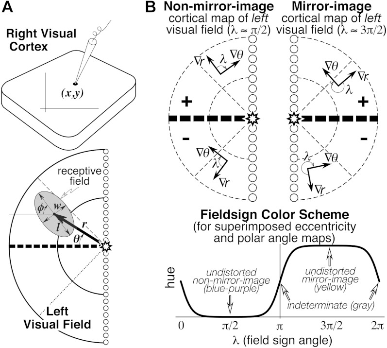
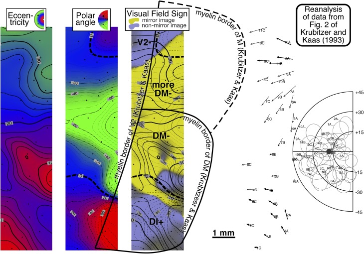
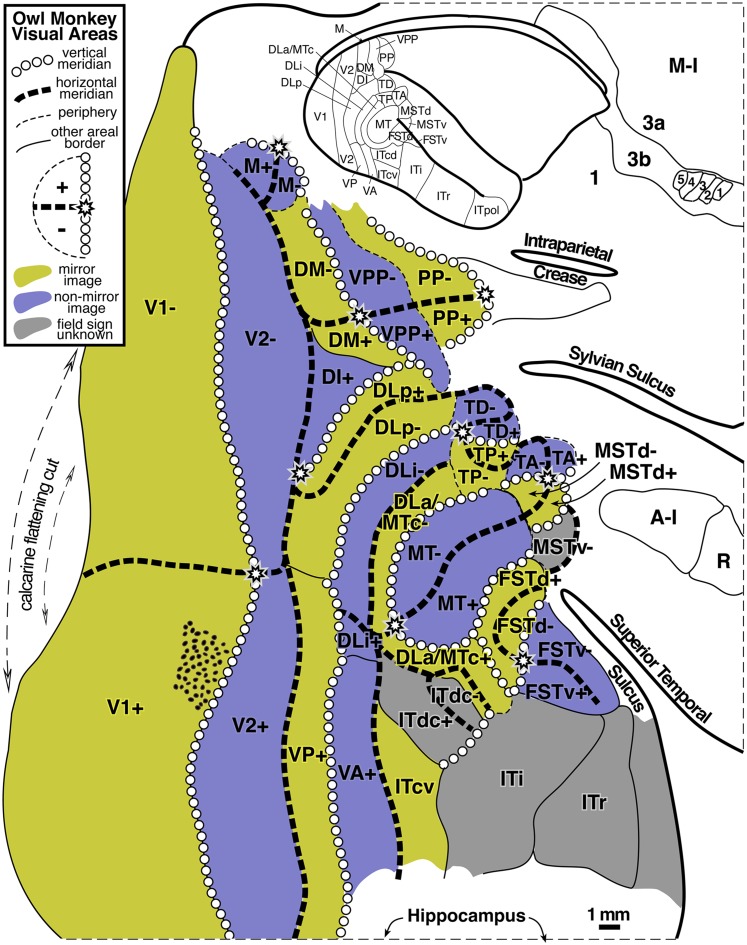
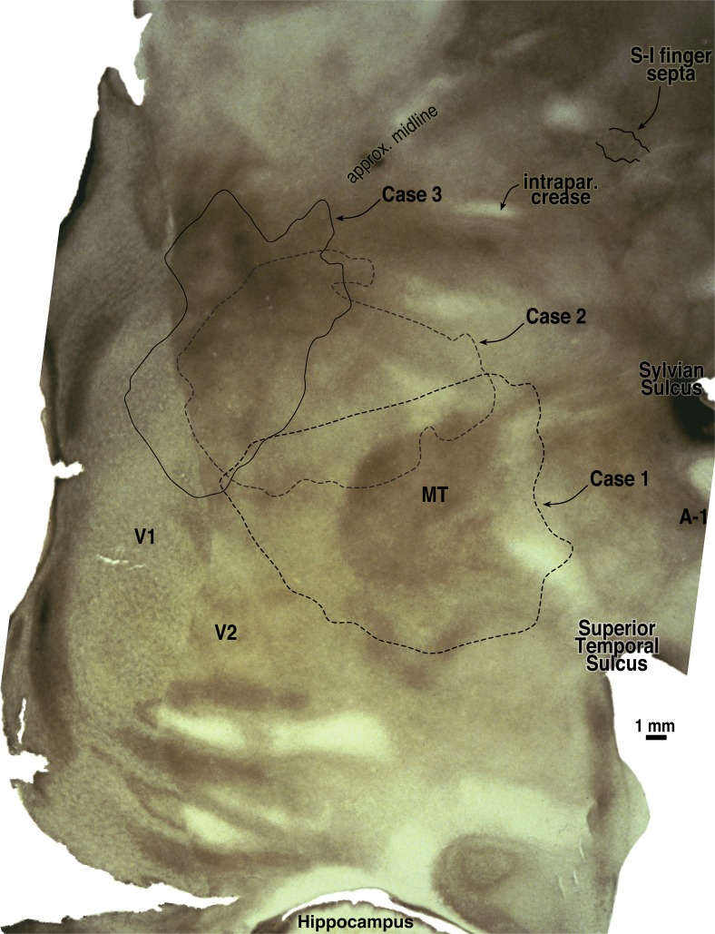
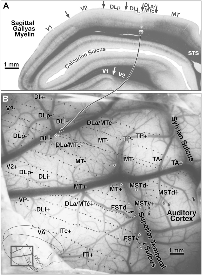
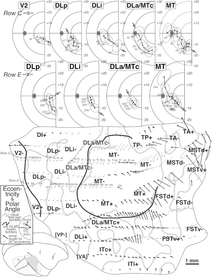
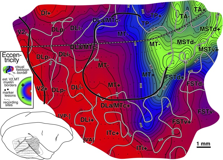
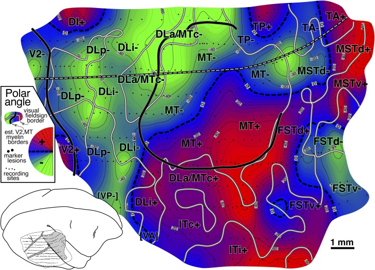
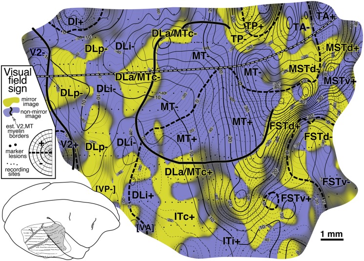
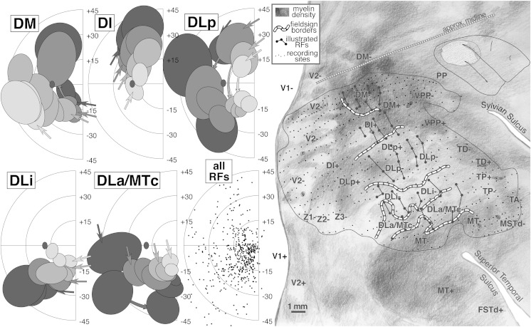
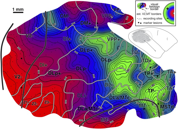
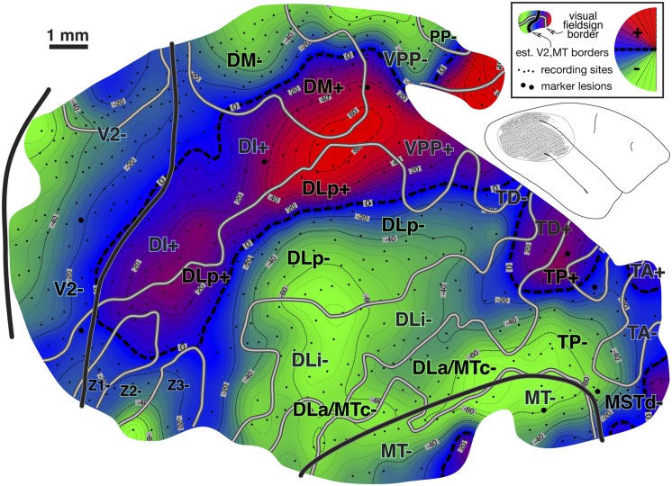
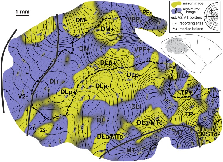
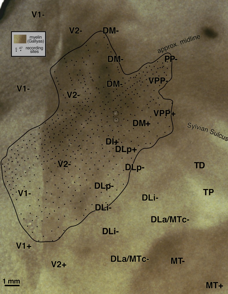
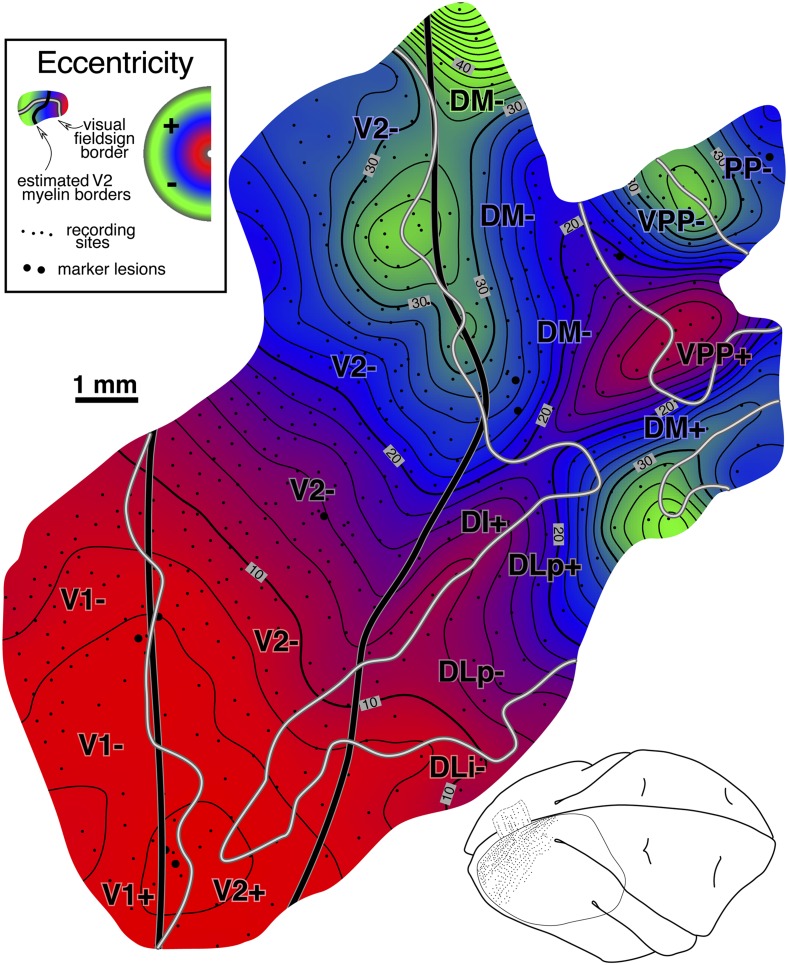
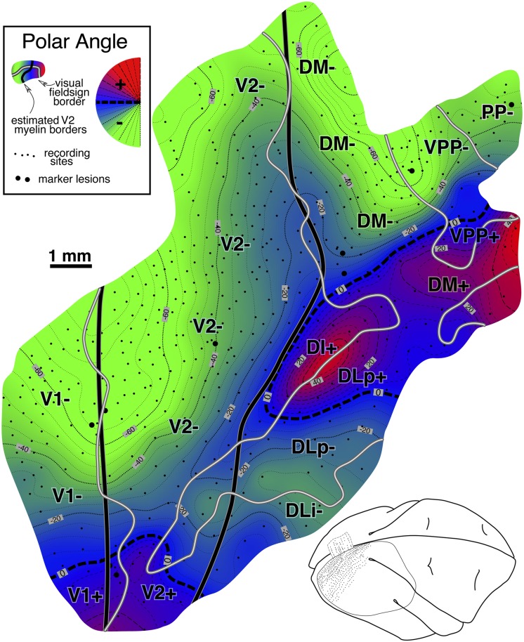
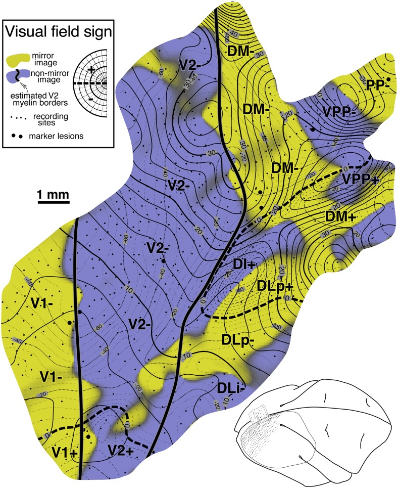
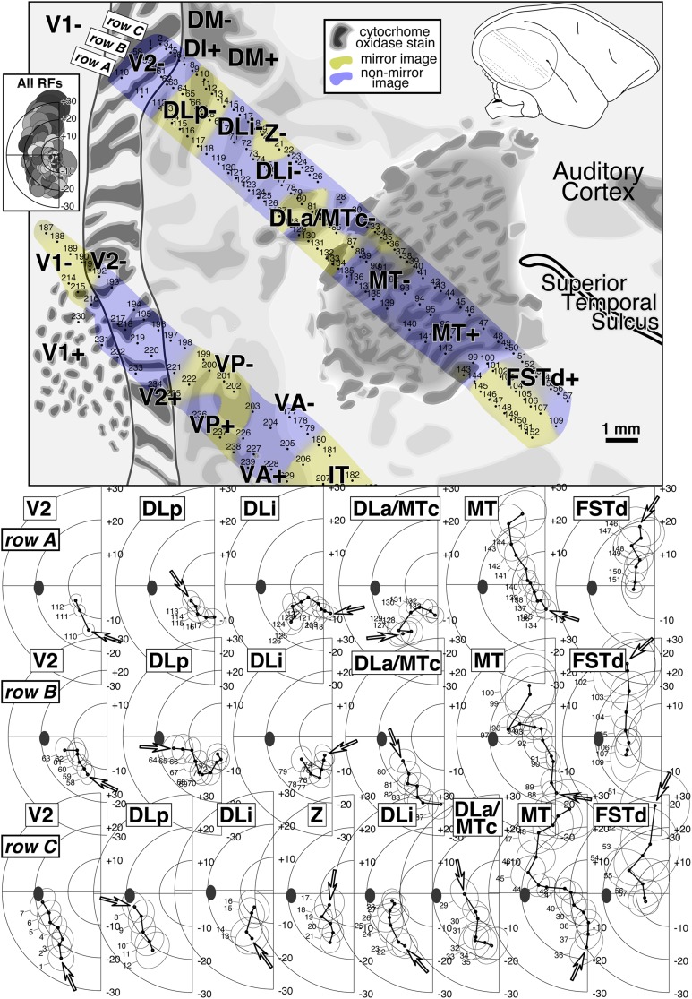
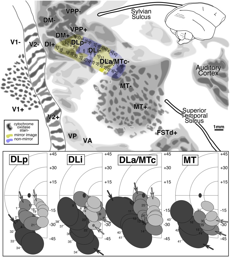
Dense retinotopy data sets were obtained by microelectrode visual receptive field mapping in dorsal and lateral visual cortex of anesthetized owl monkeys. The cortex was then physically flatmounted and stained for myelin or cytochrome oxidase. Retinotopic mapping data were digitized, interpolated to a uniform grid, analyzed using the visual field sign technique-which locally distinguishes mirror image from nonmirror image visual field representations-and correlated with the myelin or cytochrome oxidase patterns. The region between V2 (nonmirror) and MT (nonmirror) contains three areas-DLp (mirror), DLi (nonmirror), and DLa/MTc (mirror). DM (mirror) was thin anteroposteriorly, and its reduced upper field bent somewhat anteriorly away from V2. DI (nonmirror) directly adjoined V2 (nonmirror) and contained only an upper field representation that also adjoined upper field DM (mirror). Retinotopy was used to define area VPP (nonmirror), which adjoins DM anteriorly, area FSTd (mirror), which adjoins MT ventrolaterally, and TP (mirror), which adjoins MT and DLa/MTc dorsoanteriorly. There was additional retinotopic and architectonic evidence for five more subdivisions of dorsal and lateral extrastriate cortex-TA (nonmirror), MSTd (mirror), MSTv (nonmirror), FSTv (nonmirror), and PP (mirror). Our data appear quite similar to data from marmosets, though our field sign-based areal subdivisions are slightly different. The region immediately anterior to the superiorly located central lower visual field V2 varied substantially between individuals, but always contained upper fields immediately touching lower visual field V2. This region appears to vary even more between species. Though we provide a summary diagram, given within- and between-species variation, it should be regarded as a guide to parsing complex retinotopy rather than a literal representation of any individual, or as the only way to agglomerate the complex mosaic of partial upper and lower field, mirror- and nonmirror-image patches into areas.
通过在麻醉的猫头鹰猴的背侧和外侧视觉皮层进行微电极视觉感受野映射,获得了密集的视网膜拓扑数据集。然后将皮层进行物理平铺,并对髓磷脂或细胞色素氧化酶进行染色。视网膜拓扑映射数据被数字化,插值到一个均匀的网格中,使用视野标记技术进行分析——该技术在局部区分镜像和非镜像视野表示——并与髓磷脂或细胞色素氧化酶模式相关联。V2(非镜像)和MT(非镜像)之间的区域包含三个区域——DLp(镜像)、DLi(非镜像)和DLa/MTc(镜像)。DM(镜像)在前后方向上较薄,其缩小的上视野稍微向前弯曲,远离V2。DI(非镜像)直接毗邻V2(非镜像),并且只包含一个也毗邻上视野DM(镜像)的上视野表示。视网膜拓扑学被用于定义在前方毗邻DM的VPP区域(非镜像)、在腹外侧毗邻MT的FSTd区域(镜像)以及在背前方毗邻MT和DLa/MTc的TP区域(镜像)。对于背侧和外侧纹外皮层的另外五个细分区域——TA(非镜像)、MSTd(镜像)、MSTv(非镜像)、FSTv(非镜像)和PP(镜像),有更多的视网膜拓扑学和结构学证据。我们的数据看起来与狨猴的数据非常相似,尽管我们基于视野标记的区域细分略有不同。位于上方的中央下视野V2正前方的区域在个体之间差异很大,但总是包含紧邻下视野V2的上视野。这个区域在不同物种之间似乎差异更大。尽管我们提供了一个示意图,但考虑到种内和种间的变异,它应该被视为解析复杂视网膜拓扑学的指南,而不是任何个体的文字表示,也不是将部分上视野和下视野、镜像和非镜像图像斑块的复杂镶嵌整合为区域的唯一方法。