Sekino Saki, Kashiwagi Yuriko, Kanazawa Hitoshi, Takada Kazuki, Baba Takashi, Sato Seiichi, Inoue Hiroki, Kojima Masaki, Tani Katsuko
School of Life Sciences, Tokyo University of Pharmacy and Life Sciences, Hachioji, Tokyo, 192-0392, Japan.
Cell Commun Signal. 2015 Oct 1;13:41. doi: 10.1186/s12964-015-0119-5.
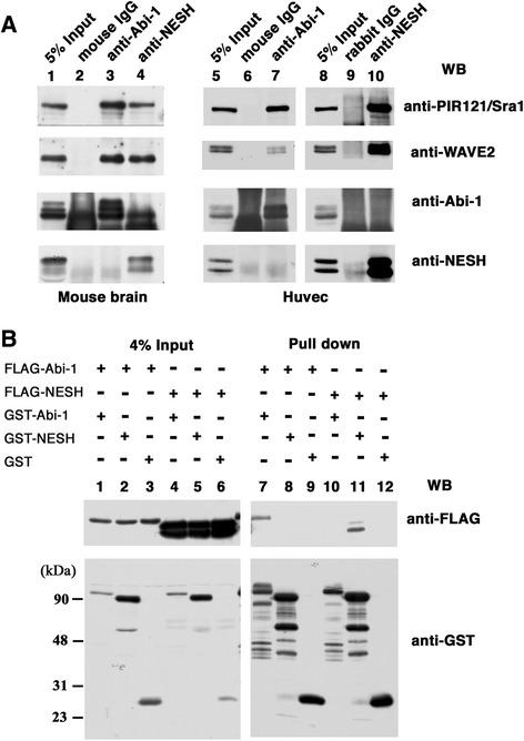
Abl interactor (Abi) family proteins play significant roles in actin cytoskeleton organization through participation in the WAVE complex. Mammals possess three Abi proteins: Abi-1, Abi-2, and NESH/Abi-3. Abi-1 and Abi-2 were originally identified as Abl tyrosine kinase-binding proteins. It has been disclosed that Abi-1 acts as a bridge between c-Abl and WAVE2, and c-Abl-mediated WAVE2 phosphorylation promotes actin remodeling. We showed previously that NESH/Abi-3 is present in the WAVE2 complex, but neither binds to c-Abl nor promotes c-Abl-mediated phosphorylation of WAVE2.
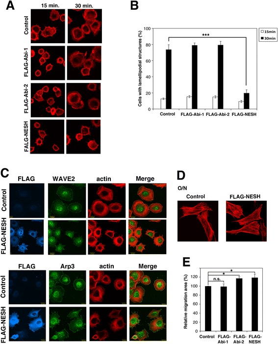
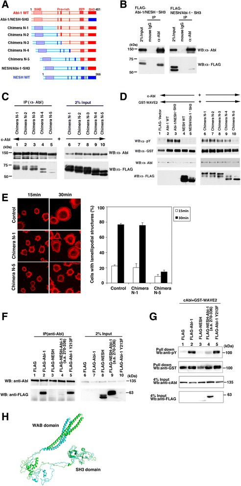
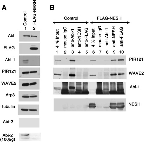
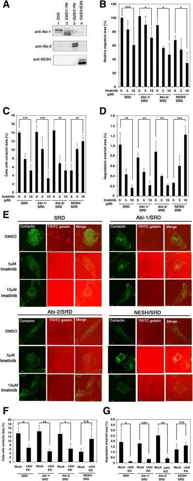
In this study, we characterized NESH/Abi-3 in more detail, and compared its properties with those of Abi-1 and Abi-2. NESH/Abi-3 was ectopically expressed in NIH3T3 cells, in which Abi-1, but not NESH/Abi-3, is expressed. The expression of NESH/Abi-3 caused degradation of endogenous Abi-1, which led to the formation of a NESH/Abi-3-based WAVE2 complex. When these cells were plated on fibronectin-coated dishes, the translocation of WAVE2 to the plasma membrane was significantly reduced and the formation of peripheral lamellipodial structures was disturbed, suggesting that the NESH/Abi-3-based WAVE2 complex was unable to help produce lamellipodial protrusions. Next, Abi-1, Abi-2, or NESH/Abi-3 was expressed in v-src-transformed NIH3T3 cells. Only in NESH/Abi-3-expressed cells did treatment with an Abl kinase inhibitor, imatinib mesylate, or siRNA-mediated knockdown of c-Abl promote the formation of invadopodia, which are ventral membrane protrusions with extracellular matrix degradation activity. Structural studies showed that a linker region between the proline-rich regions and the Src homology 3 (SH3) domain of Abi-1 is crucial for its interaction with c-Abl and c-Abl-mediated phosphorylation of WAVE2.
The NESH/Abi-3-based WAVE2 complex is functionally distinct from the Abi-1-based one, and NESH/Abi-3 may be involved in the formation of ventral protrusions under certain conditions.
Abl相互作用分子(Abi)家族蛋白通过参与WAVE复合物在肌动蛋白细胞骨架组织中发挥重要作用。哺乳动物有三种Abi蛋白:Abi-1、Abi-2和NESH/Abi-3。Abi-1和Abi-2最初被鉴定为Abl酪氨酸激酶结合蛋白。已发现Abi-1作为c-Abl与WAVE2之间的桥梁,且c-Abl介导的WAVE2磷酸化促进肌动蛋白重塑。我们之前表明NESH/Abi-3存在于WAVE2复合物中,但既不与c-Abl结合,也不促进c-Abl介导的WAVE2磷酸化。
在本研究中,我们更详细地对NESH/Abi-3进行了表征,并将其特性与Abi-1和Abi-2的特性进行了比较。NESH/Abi-3在NIH3T3细胞中异位表达,该细胞中表达Abi-1但不表达NESH/Abi-3。NESH/Abi-3的表达导致内源性Abi-1降解,这导致基于NESH/Abi-3的WAVE2复合物形成。当将这些细胞接种在纤连蛋白包被的培养皿上时,WAVE2向质膜的转位显著减少,外周板状伪足结构的形成受到干扰,这表明基于NESH/Abi-3的WAVE2复合物无法帮助产生板状伪足突起。接下来,在v-src转化的NIH3T3细胞中表达Abi-1、Abi-2或NESH/Abi-3。仅在表达NESH/Abi-3的细胞中,用Abl激酶抑制剂甲磺酸伊马替尼处理或siRNA介导的c-Abl敲低可促进侵袭性伪足的形成,侵袭性伪足是具有细胞外基质降解活性的腹侧膜突起。结构研究表明,Abi-1富含脯氨酸区域与Src同源3(SH3)结构域之间的连接区域对其与c-Abl的相互作用以及c-Abl介导的WAVE2磷酸化至关重要。
基于NESH/Abi-3的WAVE2复合物在功能上与基于Abi-1的复合物不同,并且NESH/Abi-3可能在某些条件下参与腹侧突起的形成。