Zhang H, Guo X, Zhong S, Ge T, Peng S, Yu P, Zhou Z
Jiangxi Agricultural University.
Eur J Histochem. 2015 Aug 25;59(3):2521. doi: 10.4081/ejh.2015.2521.
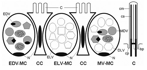
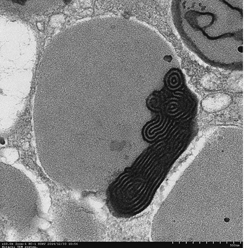
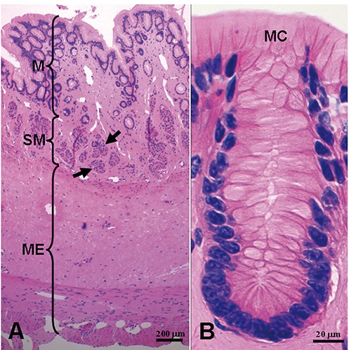
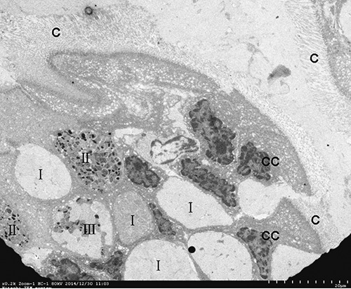
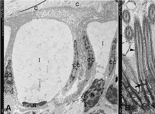
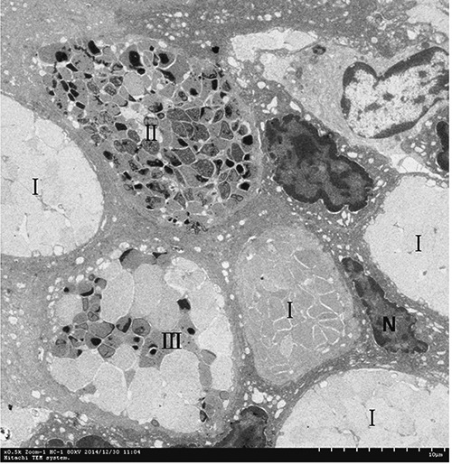
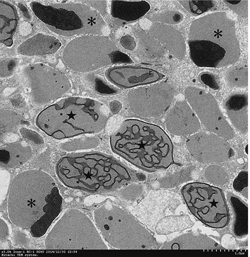
The Chinese giant salamander belongs to an old lineage of salamanders and endangered species. Many studies of breeding and disease regarding this amphibian had been implemented. However, the studies on the ultrastructure of this amphibian are rare. In this work, we provide a histological and ultrastructural investigation on posterior esophagus of Chinese giant salamander. The sections of amphibian esophagus were stained by hematoxylin & eosin (H&E). Moreover, the esophageal epithelium was observed by transmission electron microscopy (TEM). The results showed that esophageal epithelium was a single layer epithelium, which consisted of mucous cells and columnar cells. The esophageal glands were present in submucosa. The columnar cells were ciliated. According to the diverging ultrastructure of mucous vesicles, three types of mucous cells could be identified in the esophageal mucosa: i) electron-lucent vesicles mucous cell (ELV-MC); ii) electron-dense vesicles mucous cell (EDV-MC); and iii) mixed vesicles mucous cell (MV-MC).
中国大鲵属于蝾螈的古老谱系且为濒危物种。关于这种两栖动物的繁殖和疾病已有许多研究。然而,对这种两栖动物超微结构的研究却很罕见。在这项工作中,我们对中国大鲵食管后部进行了组织学和超微结构研究。两栖动物食管切片用苏木精和伊红(H&E)染色。此外,通过透射电子显微镜(TEM)观察食管上皮。结果表明,食管上皮为单层上皮,由黏液细胞和柱状细胞组成。食管腺存在于黏膜下层。柱状细胞有纤毛。根据黏液小泡的不同超微结构,在食管黏膜中可识别出三种类型的黏液细胞:i)电子透明小泡黏液细胞(ELV-MC);ii)电子致密小泡黏液细胞(EDV-MC);iii)混合小泡黏液细胞(MV-MC)。