Lok Sjoukje I, de Jonge Nicolaas, van Kuik Joyce, van Geffen Ankie J P, Huibers Manon M H, van der Weide Petra, Siera Erica, Winkens Bjorn, Doevendans Pieter A, de Weger Roel A, da Costa Martins Paula A
Department of Cardiology, University Medical Center, Utrecht, the Netherlands; Department of Pathology, University Medical Center, Utrecht, the Netherlands.
Department of Cardiology, University Medical Center, Utrecht, the Netherlands.
PLoS One. 2015 Oct 2;10(10):e0136404. doi: 10.1371/journal.pone.0136404. eCollection 2015.
Pulsatile flow left ventricular assist devices (pf-LVADs) are being replaced by continuous flow LVADs (cf-LVADs) in patients with end-stage heart failure (HF). MicroRNAs (miRs) play an important role in the onset and progression of HF. Our aim was to analyze cardiac miR expression patterns associated with each type of device, to analyze differences in the regulation of the induced cardiac changes.
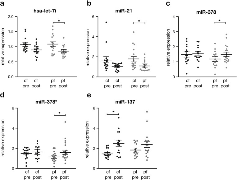
Twenty-six miRs were selected (based on micro-array data and literature studies) and validated in myocardial tissue before and after pf- (n = 17) and cf-LVAD (n = 17) support. Of these, 5 miRs displayed a similar expression pattern among the devices (miR-129*, miR-146a, miR-155, miR-221, miR-222), whereas others only changed significantly during pf-LVAD (miR-let-7i, miR-21, miR-378, miR-378*) or cf-LVAD support (miR-137). In addition, 4 miRs were investigated in plasma of cf-LVAD supported patients (n = 18) and healthy controls (n = 10). Circulating miR-21 decreased at 1, 3, and 6 months after LVAD implantation. MiR-146a, miR-221 and miR-222 showed a fluctuating time pattern post-LVAD.
Our data show a different miR expression pattern after LVAD support, suggesting that differentially expressed miRs are partially responsible for the cardiac morphological and functional changes observed after support. However, the miR expression patterns do not seem to significantly differ between pf- and cf-LVAD implying that most cardiac changes or clinical outcomes specific to each device do not relate to differences in miR expression levels.
在终末期心力衰竭(HF)患者中,搏动血流左心室辅助装置(pf-LVADs)正被连续血流左心室辅助装置(cf-LVADs)所取代。微小RNA(miRs)在HF的发生和发展中起重要作用。我们的目的是分析与每种装置相关的心脏miR表达模式,分析诱导的心脏变化调节方面的差异。
基于微阵列数据和文献研究选择了26种miRs,并在pf-LVAD(n = 17)和cf-LVAD(n = 17)支持前后的心肌组织中进行验证。其中,5种miRs在各装置间表现出相似的表达模式(miR-129*、miR-146a、miR-155、miR-221、miR-222),而其他miRs仅在pf-LVAD(miR-let-7i、miR-21、miR-378、miR-378*)或cf-LVAD支持期间(miR-137)有显著变化。此外,在cf-LVAD支持患者(n = 18)和健康对照(n = 10)的血浆中研究了4种miRs。LVAD植入后1、3和6个月,循环miR-21降低。miR-146a、miR-221和miR-222在LVAD后呈现波动的时间模式。
我们的数据显示LVAD支持后miR表达模式不同,表明差异表达的miRs部分导致了支持后观察到的心脏形态和功能变化。然而,pf-LVAD和cf-LVAD之间的miR表达模式似乎没有显著差异,这意味着每种装置特有的大多数心脏变化或临床结果与miR表达水平的差异无关。