Liu Yu, Fite Brett Z, Mahakian Lisa M, Johnson Sarah M, Larrat Benoit, Dumont Erik, Ferrara Katherine W
Department of Biomedical Engineering, University of California Davis, Davis, CA, 95616, United States of America.
UNité d'Imagerie par Résonance Magnétique et Spectroscopie, NeuroSpin, CEA, Gif Sur Yvette, France.
PLoS One. 2015 Oct 6;10(10):e0139667. doi: 10.1371/journal.pone.0139667. eCollection 2015.
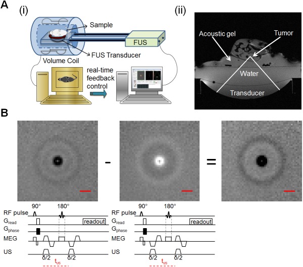
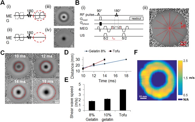
Manual palpation is a common and very informative diagnostic tool based on estimation of changes in the stiffness of tissues that result from pathology. In the case of a small lesion or a lesion that is located deep within the body, it is difficult for changes in mechanical properties of tissue to be detected or evaluated via palpation. Furthermore, palpation is non-quantitative and cannot be used to localize the lesion. Magnetic Resonance-guided Focused Ultrasound (MRgFUS) can also be used to evaluate the properties of biological tissues non-invasively. In this study, an MRgFUS system combines high field (7T) MR and 3 MHz focused ultrasound to provide high resolution MR imaging and a small ultrasonic interrogation region (~0.5 x 0.5 x 2 mm), as compared with current clinical systems. MR-Acoustic Radiation Force Imaging (MR-ARFI) provides a reliable and efficient method for beam localization by detecting micron-scale displacements induced by ultrasound mechanical forces. The first aim of this study is to develop a sequence that can concurrently quantify acoustic radiation force displacements and image the resulting transient shear wave. Our motivation in combining these two measurements is to develop a technique that can rapidly provide both ARFI and shear wave velocity estimation data, making it suitable for use in interventional radiology. Secondly, we validate this sequence in vivo by estimating the displacement before and after high intensity focused ultrasound (HIFU) ablation, and we validate the shear wave velocity in vitro using tissue-mimicking gelatin and tofu phantoms. Such rapid acquisitions are especially useful in interventional radiology applications where minimizing scan time is highly desirable.
手动触诊是一种常见且信息丰富的诊断工具,基于对因病理变化导致的组织硬度改变的评估。对于小病变或位于身体深部的病变,很难通过触诊检测或评估组织力学性能的变化。此外,触诊是非定量的,不能用于定位病变。磁共振引导聚焦超声(MRgFUS)也可用于无创评估生物组织的特性。在本研究中,一个MRgFUS系统结合了高场(7T)磁共振和3MHz聚焦超声,与当前临床系统相比,可提供高分辨率磁共振成像和一个小的超声询问区域(约0.5×0.5×2mm)。磁共振声辐射力成像(MR-ARFI)通过检测超声机械力引起的微米级位移,为束定位提供了一种可靠且高效的方法。本研究的首要目标是开发一种序列,能够同时量化声辐射力位移并对产生的瞬态剪切波进行成像。我们将这两种测量相结合的动机是开发一种技术,能够快速提供ARFI和剪切波速度估计数据,使其适用于介入放射学。其次,我们通过估计高强度聚焦超声(HIFU)消融前后的位移在体内验证该序列,并使用组织模拟明胶和豆腐体模在体外验证剪切波速度。这种快速采集在介入放射学应用中特别有用,因为在这些应用中非常希望尽量缩短扫描时间。