Hersh David S, Anastasiadis Pavlos, Mohammadabadi Ali, Nguyen Ben A, Guo Sijia, Winkles Jeffrey A, Kim Anthony J, Gullapalli Rao, Keller Asaf, Frenkel Victor, Woodworth Graeme F
Department of Neurosurgery, University of Maryland School of Medicine, Baltimore, Maryland, United States of America.
Department of Diagnostic Radiology and Nuclear Medicine, University of Maryland School of Medicine, Baltimore, Maryland, United States of America.
PLoS One. 2018 Feb 7;13(2):e0192240. doi: 10.1371/journal.pone.0192240. eCollection 2018.
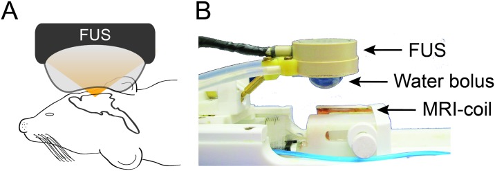
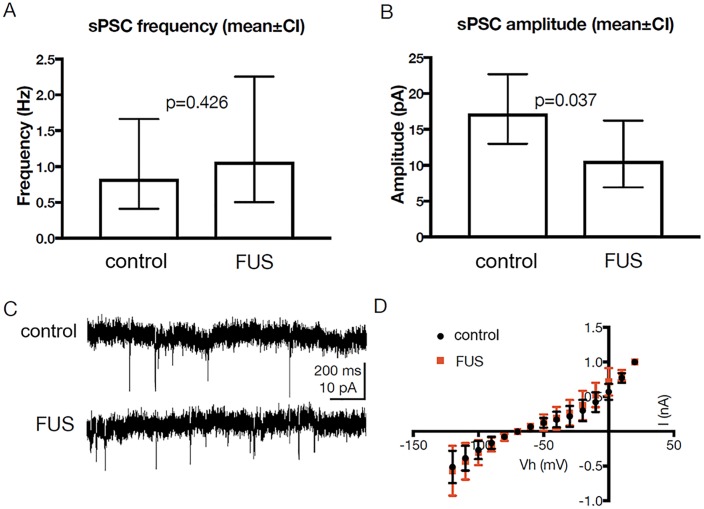
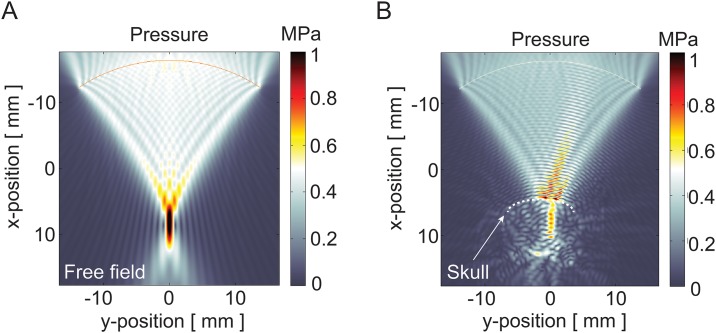
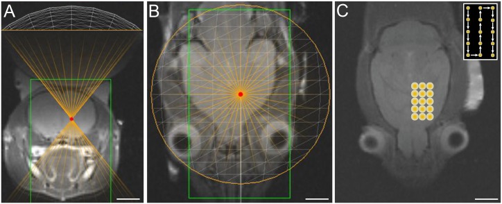
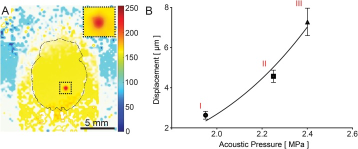
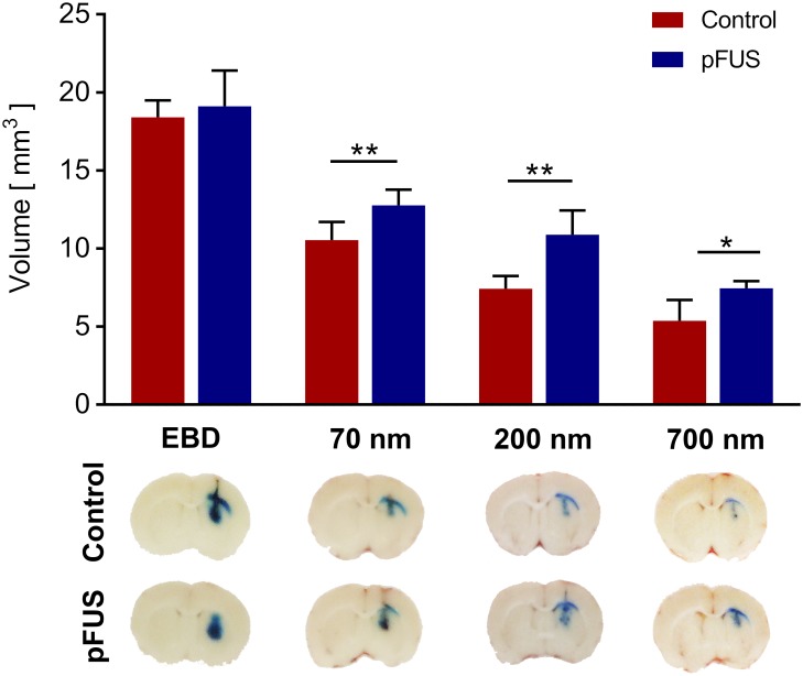
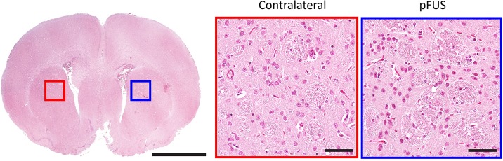
Generating spatially controlled, non-destructive changes in the interstitial spaces of the brain has a host of potential clinical applications, including enhancing the delivery of therapeutics, modulating biological features within the tissue microenvironment, altering fluid and pressure dynamics, and increasing the clearance of toxins, such as plaques found in Alzheimer's disease. Recently we demonstrated that ultrasound can non-destructively enlarge the interstitial spaces of the brain ex vivo. The goal of the current study was to determine whether these effects could be reproduced in the living brain using non-invasive, transcranial MRI-guided focused ultrasound (MRgFUS). The left striatum of healthy rats was treated using MRgFUS. Computer simulations facilitated treatment planning, and targeting was validated using MRI acoustic radiation force impulse imaging. Following MRgFUS treatments, Evans blue dye or nanoparticle probes were infused to assess changes in the interstitial space. In MRgFUS-treated animals, enhanced dispersion was observed compared to controls for 70 nm (12.8 ± 0.9 mm3 vs. 10.6 ± 1.0 mm3, p = 0.01), 200 nm (10.9 ± 1.4 mm3 vs. 7.4 ± 0.7 mm3, p = 0.01) and 700 nm (7.5 ± 0.4 mm3 vs. 5.4 ± 1.2 mm3, p = 0.02) nanoparticles, indicating enlargement of the interstitial spaces. No evidence of significant histological or electrophysiological injury was identified. These findings suggest that transcranial ultrasound can safely and effectively modulate the brain interstitium and increase the dispersion of large therapeutic entities such as particulate drug carriers or modified viruses. This has the potential to expand the therapeutic uses of MRgFUS.
在脑间质空间中产生空间可控的、非破坏性的变化具有许多潜在的临床应用,包括增强治疗药物的递送、调节组织微环境中的生物学特性、改变流体和压力动力学,以及增加毒素的清除,如阿尔茨海默病中发现的斑块。最近我们证明,超声可以在体外非破坏性地扩大脑间质空间。本研究的目的是确定是否可以使用非侵入性的、经颅MRI引导聚焦超声(MRgFUS)在活体脑中再现这些效应。使用MRgFUS对健康大鼠的左侧纹状体进行治疗。计算机模拟有助于治疗计划的制定,并使用MRI声辐射力脉冲成像验证靶向定位。在MRgFUS治疗后,注入伊文思蓝染料或纳米颗粒探针以评估间质空间的变化。在接受MRgFUS治疗的动物中,观察到70 nm(12.8±0.9 mm³对10.6±1.0 mm³,p = 0.01)、200 nm(10.9±1.4 mm³对7.4±0.7 mm³,p = 0.01)和700 nm(7.5±0.4 mm³对5.4±1.2 mm³,p = 0.02)纳米颗粒的扩散增强,表明间质空间扩大。未发现明显的组织学或电生理损伤证据。这些发现表明,经颅超声可以安全有效地调节脑间质,并增加大型治疗实体如颗粒药物载体或修饰病毒的扩散。这有可能扩大MRgFUS的治疗用途。